
Page 108 - Read Online
P. 108
Page 4 of 15 Mathew et al. Vessel Plus 2020;4:11 I http://dx.doi.org/10.20517/2574-1209.2019.35
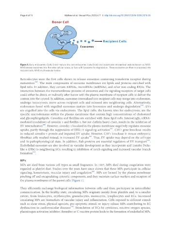
Figure 1. Early endosomes (Early Endo) mature into late endosomes (Late Endo) and accumulate intraluminal vesicles known as MVB.
MVB release exosomes into the extra cellular space, or fuse with lysosome for degradation. These exosomes are then incorporated into
recipient cells. MVB: multivesicular bodies
Reticulocytes were the first cells shown to release exosomes containing transferrin receptor during
[38]
maturation . The main components of exosome membranes are lipids and proteins enriched with
lipid rafts. In addition, they contain mRNAs, microRNAs (miRNAs), and other non-coding RNAs. The
interaction between the transmembrane proteins of exosomes and the signaling receptors of target cells
could either be direct, or indirectly after fusion with the plasma membrane of recipient cells to deliver the
content into the cytosol. In addition, exosomes internalized into recipient cells may merge into endosomes,
undergo transcytosis, move across recipient cells and released into neighboring cells. Alternatively,
[39]
endosomes fused with engulfed exosomes mature into lysosomes and undergo degradation . EVs
are engulfed into the cells via endocytosis. The lipid rafts, the known sites for endocytosis, are the
specific microdomains within the plasma membrane that contain high concentrations of cholesterol
and glycosphingolipids. Caveolins and flotillins are enriched with these lipid rafts. Interestingly, siRNA-
mediated knockdown of caveolin-1 and flotillin-1, but not clathrin heavy chain, results in the inhibition of
[40]
EV internalization . However, caveolin-1 localized in the plasma membrane negatively regulates exosome
[41]
uptake, partly through the suppression of ERK1/2 signaling activation . CAV1 gene knockout results
in reduced caveolin-1 protein and impaired EV uptake. However, CAV1 knockout in mouse embryonic
fibroblast cells resulted instead, in increased EV uptake . Thus, EV uptake may depend on the cell type
[42]
[43]
and its pathophysiological state. In addition, Rab proteins are essential regulators of EV transport .
Endothelial exosomes are also involved in vascular development as they incorporate and transfer Delta-
like 4 (Dll4) to neighboring ECs, resulting in inhibition of notch signaling and increased vascular branch
[44]
formation .
MPs
MPs are shed from various cell types as small fragments. In 1967, MPs shed during coagulation were
regarded as platelet dust. Studies over the years have since shown that these MPs participate in cellular
[45]
signaling, homeostasis, vascular injury and coagulation . MPs are formed by the plasma membrane
pinching off and encapsulating cytosolic components, and they maintain surface markers and receptors of
the plasma membranes of the parent cells [Figure 2].
They efficiently exchange biological information between cells and thus, participate in intercellular
communication. In the healthy state, circulating MPs originate mainly from platelets and, to a smaller
extent, from leukocytes, erythrocytes, granulocytes, monocytes, lymphocytes and ECs. Increased
circulating MPs are biomarkers of vascular injury and inflammation. Cells exposed to different stimuli
such as shear stress, physical agonists, pro-apoptotic stimuli or injury release MPs contributing to EC
dysfunction in cardiovascular diseases . Stimulation of ECs by cytokines, reactive oxygen species,
[46]
plasminogen activation inhibitor, thrombin or C-reactive protein leads to the formation of endothelial MPs.