
Page 106 - Read Online
P. 106
Page 2 of 15 Mathew et al. Vessel Plus 2020;4:11 I http://dx.doi.org/10.20517/2574-1209.2019.35
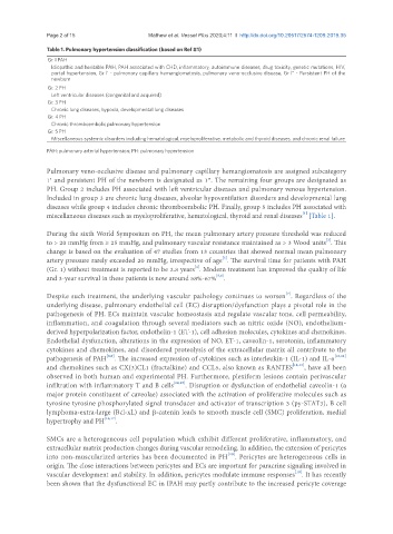
Table 1. Pulmonary hypertension classification (based on Ref #1)
Gr. l PAH
Idiopathic and heritable PAH, PAH associated with CHD, inflammatory, autoimmune diseases, drug toxicity, genetic mutations, HIV,
portal hypertension, Gr l’ - pulmonary capillary hemangiomatosis, pulmonary veno- occlusive disease, Gr l” - Persistent PH of the
newborn
Gr. 2 PH
Left ventricular diseases (congenital and acquired)
Gr. 3 PH
Chronic lung diseases, hypoxia, developmental lung diseases
Gr. 4 PH
Chronic thromboembolic pulmonary hypertension
Gr. 5 PH
Miscellaneous systemic disorders including hematological, myeloproliferative, metabolic and thyroid diseases, and chronic renal failure
PAH: pulmonary arterial hypertension; PH: pulmonary hypertension
Pulmonary veno-occlusive disease and pulmonary capillary hemangiomatosis are assigned subcategory
1′ and persistent PH of the newborn is designated as 1″. The remaining four groups are designated as
PH. Group 2 includes PH associated with left ventricular diseases and pulmonary venous hypertension.
Included in group 3 are chronic lung diseases, alveolar hypoventilation disorders and developmental lung
diseases while group 4 includes chronic thromboembolic PH. Finally, group 5 includes PH associated with
[1]
miscellaneous diseases such as myeloproliferative, hematological, thyroid and renal diseases [Table 1].
During the sixth World Symposium on PH, the mean pulmonary artery pressure threshold was reduced
[2]
to > 20 mmHg from ≥ 25 mmHg, and pulmonary vascular resistance maintained as > 3 Wood units . This
change is based on the evaluation of 47 studies from 13 countries that showed normal mean pulmonary
[3]
artery pressure rarely exceeded 20 mmHg, irrespective of age . The survival time for patients with PAH
[4]
(Gr. 1) without treatment is reported to be 2.8 years . Modern treatment has improved the quality of life
[5,6]
and 3-year survival in these patients is now around 58%-67% .
Despite such treatment, the underlying vascular pathology continues to worsen . Regardless of the
[7]
underlying disease, pulmonary endothelial cell (EC) disruption/dysfunction plays a pivotal role in the
pathogenesis of PH. ECs maintain vascular homeostasis and regulate vascular tone, cell permeability,
inflammation, and coagulation through several mediators such as nitric oxide (NO), endothelium-
derived hyperpolarization factor, endothelin-1 (ET-1), cell adhesion molecules, cytokines and chemokines.
Endothelial dysfunction, alterations in the expression of NO, ET-1, caveolin-1, serotonin, inflammatory
cytokines and chemokines, and disordered proteolysis of the extracellular matrix all contribute to the
[8,9]
pathogenesis of PAH . The increased expression of cytokines such as interleukin-1 (IL-1) and IL-6 [10,11]
and chemokines such as CX(3)CL1 (fractalkine) and CCL5, also known as RANTES [12,13] , have all been
observed in both human and experimental PH. Furthermore, plexiform lesions contain perivascular
infiltration with inflammatory T and B cells [14,15] . Disruption or dysfunction of endothelial caveolin-1 (a
major protein constituent of caveolae) associated with the activation of proliferative molecules such as
tyrosine tyrosine phosphorylated signal transducer and activator of transcription 3 (py-STAT3), B cell
lymphoma-extra-large (Bcl-xL) and β-catenin leads to smooth muscle cell (SMC) proliferation, medial
hypertrophy and PH [16,17] .
SMCs are a heterogeneous cell population which exhibit different proliferative, inflammatory, and
extracellular matrix production changes during vascular remodeling. In addition, the extension of pericytes
[18]
into non-muscularized arteries has been documented in PH . Pericytes are heterogeneous cells in
origin. The close interactions between pericytes and ECs are important for paracrine signaling involved in
vascular development and stability. In addition, pericytes modulate immune responses . It has recently
[19]
been shown that the dysfunctional EC in IPAH may partly contribute to the increased pericyte coverage