
Page 394 - Read Online
P. 394
Page 8 of 13 Iqbal et al. Vessel Plus 2019;3:40 I http://dx.doi.org/10.20517/2574-1209.2019.28
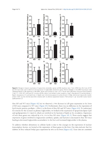
Figure 5. Changes in hepatic expression of triglycerides metabolism genes in RORγ knockout mice. Total mRNA from the livers of WT
and KO mice were used to determine the expression of triglycerides metabolism genes. Relative changes in the mRNA expression were
calculated based on the expression of 18 sRNA. Values were plotted as mean ± SD. P values were calculated using two-tailed Student’s
t test. *P < 0.05. WT: wild type; KO: knockout; RORγ: retinoic acid-related orphan receptor γ; Dgat1: diacylglycerol O-acyltransferase 1;
Dgat2: diacylglycerol O-acyltransferase 2; Mgat2: acyl CoA:monoacylglycerol acyltransferase 2; Mttp: microsomal triglyceride transfer
protein; Vldlr: very low-density lipoprotein receptor; Ldlr: low-density lipoprotein receptor; Plin1: perilipin 1; Lpl: lipoprotein lipase; Apoc2:
apolipoprotein C2
Rorγ KO and WT mice [Figure 5E] but we observed a 79% decrease in Ldlr gene expression in the livers
of KO mice compared to WT mice [Figure 5F]. Furthermore, there was no difference in the expression of
lipid droplet protein, perilipin 1 (Plin1), in the livers of Rorγ KO and WT mice [Figure 5G]. To understand
the reasons for the increase in plasma triglycerides, we looked at the expression of lipoprotein lipase (Lpl)
and apolipoprotein C2 (ApoC2), which are involved in the lipolysis of lipids in the circulation. Expression
of both these genes was reduced by 67%-71% in Rorγ KO mice [Figure 5H, I]. These results suggest that
expression of genes involved in triglycerides synthesis, uptake, and lipolysis is decreased in Rorγ KO mice
leading to decreased triglycerides accumulation in the liver and increased triglycerides in the plasma.
To address whether alterations in cellular lipids is due to the changes in the expression of nuclear
transcription factors, we looked at the expression of these genes in the liver. Our data indicate that the
deletion of Rorγ reduced Srebp2 gene expression by 80% in the livers [Figure 6A]. These data are consistent