
Page 374 - Read Online
P. 374
Page 6 of 12 Mestres et al. Vessel Plus 2019;3:38 I http://dx.doi.org/10.20517/2574-1209.2019.20
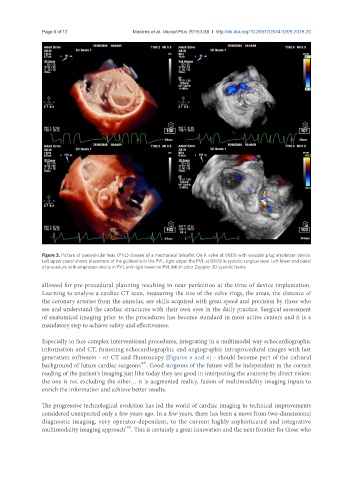
Figure 3. Picture of paravalvular leak (PVL) closure of a mechanical bileaflet On-X valve at 0800 with vascular plug amplatzer device.
Left upper panel shows placement of the guidewire in the PVL, right upper the PVL at 0800 in systolic surgical view. Left lower end panel
of procedure with amplatzer device in PVL and right lower no PVL left in color Doppler 3D systolic frame
allowed for pre-procedural planning resulting in near perfection at the time of device implantation.
Learning to analyze a cardiac CT scan, measuring the size of the valve rings, the areas, the distance of
the coronary arteries from the annulus, are skills acquired with great speed and precision by those who
see and understand the cardiac structures with their own eyes in the daily practice. Surgical assessment
of anatomical imaging prior to the procedures has become standard in most active centers and it is a
mandatory step to achieve safety and effectiveness.
Especially to face complex interventional procedures, integrating in a multimodal way echocardiographic
information and CT, fusioning echocardiographic and angiographic intraprocedural images with last
generation softwares - or CT and fluoroscopy [Figures 5 and 6] - should become part of the cultural
[47]
background of future cardiac surgeons . Good surgeons of the future will be independent in the correct
reading of the patient’s imaging just like today they are good in interpreting the anatomy by direct vision:
the one is not excluding the other… it is augmented reality, fusion of multimodality imaging inputs to
enrich the information and achieve better results.
The progressive technological evolution has led the world of cardiac imaging to technical improvements
considered unexpected only a few years ago. In a few years, there has been a move from two-dimensional
diagnostic imaging, very operator-dependent, to the current highly sophisticated and integrative
[48]
multimodality imaging approach . This is certainly a great innovation and the next frontier for those who