
Page 373 - Read Online
P. 373
Mestres et al. Vessel Plus 2019;3:38 I http://dx.doi.org/10.20517/2574-1209.2019.20 Page 5 of 12
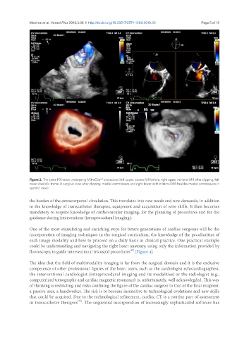
Figure 2. The same P3 lesion undergoing MitraClip™ procedure (left upper: severe MR before, right upper minimal MR after clipping, left
lower diastolic frame in surgical view after clipping, medial commissure and right lower with minimal MR besides medial commissure in
systolic view)
the burden of the extracorporeal circulation. This translates into new needs and new demands, in addition
to the knowledge of transcatheter therapies, equipment and acquisition of wire skills. It then becomes
mandatory to acquire knowledge of cardiovascular imaging, for the planning of procedures and for the
guidance during interventions (intraprocedural imaging).
One of the most stimulating and enriching steps for future generations of cardiac surgeons will be the
incorporation of imaging techniques in the surgical curriculum, the knowledge of the peculiarities of
each image modality and how to proceed on a daily basis in clinical practice. One practical example
could be understanding and navigating the right heart anatomy, using only the information provided by
[45]
fluoroscopy, to guide interventional tricuspid procedures [Figure 4].
The idea that the field of multimodality imaging is far from the surgical domain and it is the exclusive
competence of other professional figures of the heart team, such as the cardiologist echocardiographist,
the interventional cardiologist (intraprocedural imaging and its modalities) or the radiologist (e.g.,
computerized tomography and cardiac magnetic resonance) is, unfortunately, well acknowlegded. This way
of thinking is restricting and risks confining the figure of the cardiac surgeon to that of the final recipient,
a passive user, a handworker. The risk is to become insensitive to technological evolutions and new skills
that could be acquired. Due to the technological refinement, cardiac CT is a routine part of assessment
[46]
in transcatheter therapies . The sequential incorporation of increasingly sophisticated software has