
Page 372 - Read Online
P. 372
Page 4 of 12 Mestres et al. Vessel Plus 2019;3:38 I http://dx.doi.org/10.20517/2574-1209.2019.20
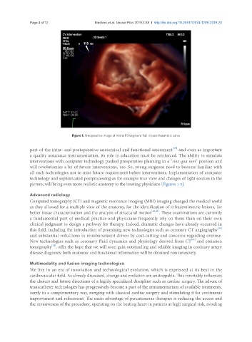
Figure 1. Preoperative image of mitral P3 segment flail in postrheumatic valve
[39]
part of the intra- and postoperative anatomical and functional assessment and even as important
a quality assurance instrumentation, its role in education must be reinforced. The ability to simulate
interventions with computer technology pushed preoperative planning in a “sine qua non” position and
will revolutionize a lot of future interventions, too. So, young surgeons need to become familiar with
all such technologies not to miss future requirement before interventions. Implementation of computer
technology and sophisticated postprocessing as for example true view and changes of light sources in the
picture, will bring even more realistic anatomy to the treating physicians [Figures 1-3].
Advanced radiology
Computed tomography (CT) and magnetic resonance imaging (MRI) imaging changed the medical world
as they allowed for a multiple view of the anatomy, for the identification of infracentimetric lesions, for
better tissue characterization and the analysis of structural motion [40,41] . These examinations are currently
a fundamental part of medical practice and physicians frequently rely on them than on their own
clinical judgment to design a pathway for therapy. Indeed, dramatic changes have already occurred in
[42]
this field, including the introduction of promising new technologies such as coronary CT angiography
and substantial reductions in reimbursement driven by cost-cutting and concerns regarding overuse.
[43]
New technologies such as coronary fluid dynamics and physiology derived from CT and emission
[44]
tomography , offer the hope that we will soon gain outstanding and reliable imaging in coronary artery
disease diagnosis: both anatomic and functional information will be obtained non invasively.
Multimodality and fusion imaging technologies
We live in an era of innovation and technological evolution, which is expressed at its best in the
cardiovascular field. As already discussed, change and evolution are unstoppable. This inevitably influences
the choices and future directions of a highly specialized discipline such as cardiac surgery. The advent of
transcatheter technologies has progressively become a part of the armamentarium of available treatments,
surely in a complementary way, merging with classical cardiac surgery and stimulating it for continuous
improvement and refinement. The main advantage of percutaneous therapies is reducing the access and
the invasiveness of the procedure, operating on the beating heart in patients at high surgical risk, avoiding