
Page 127 - Read Online
P. 127
Paraggio et al. Vessel Plus 2019;3:12 I http://dx.doi.org/10.20517/2574-1209.2018.72 Page 9 of 11
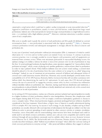
Table 4. Ellis classification of coronary perforation [30]
Ellis class Definition
I Extraluminal crater without extravasation
II Pericardial or myocardial blushing
III Width of perforation ≥ 1 mm with contrast streaming and cavity spilling
[29]
catastrophic complication which could lead to sudden cardiac tamponade or acute myocardial infarction .
Aggressive predilation or postdilation, usually in more calcified lesions, could determine large vessel
perforations. Indeed, one of the most predictive factors for large vessel perforation is a high balloon-to-artery
[29]
ratio > 1.2 combined with a high inflation pressure . Moreover, rotational aterectomy is another common
cause of such complication.
Ellis score is usually used to grade the severity of such perforation and Ellis grade III (defined as contrast
[30]
extravasation from > 1 mm perforation) is associated with the highest mortality [Table 4]. However,
coronary perforation severity and subsequent management is strongly affected by clinical scenario and
perforation site.
In any case of proximal vessel perforation without jet extravasation (Ellis 1), treatment is limited to careful
observation for 15-30 min and no further actions are needed if does not increase. In contrast to previous
common practice, now is strongly avoided to reverse heparin with protamine until all equipments are
removed from coronary artery. When even minimum pericardial or myocardial blushing occurs, the
optimal strategy is to inflate a balloon for about 10 min at low pressure over the site of perforation to stop
the flow. To avoid prolonged ischemia during this phase, some operators suggested the “microcatheter distal
perfusion technique”, which consist of placing over another guidewire a microcatheter distal to perforation
in order to inject patient’s blood. When balloon inflation does not achieve vessel sealing, a covered stent
should be placed in the site of perforation from the same catheter or by using the so-called “ping-pong
technique”. Indeed, in case of consistent jet extravasation, removal of balloon and subsequent deliver of
covered stent could determine massive blood loss. Moreover, only recently developed covered stents shows
compatibility with 6F GC (PK Papyrus, Biotronik, Berlin, Germany) but they still not allow coexistence with
balloon shaft, thus determining the need for GC replacement. In such cases, this CTO derived technique
conveys the use of a second GC to engage the coronary ostium while the first is withdraw few millimeters
without deinflate balloon at perforation site. Therefore, during rapid balloon deinflation and reinflation, a
second guidewire is placed distally. Such balloon is finally deinflated and retrieved only while covered stent
is delivered at the site of perforation.
In the setting of CTO interventions, routine use of heavier, polymer jacketed and tapered guidewires results
in a higher incidence of distal perforation related to wire manipulation. However, this complication could
even happen during daily PCI when a CTO (or simply a workhorse hydrophilic) guidewire is chosen to
cross a complex lesion. When distal perforation happen, proximal prolonged balloon inflation should be
considered as first line treatment. Nevertheless, a higher dose of heparin is usually administered during
CTO or complex PCI thus lowering the success of such strategy. In past decades, complete sealing of distal
perforation was usually obtained with local injection of subcutaneous fat through an inflated over-the-wire
balloon in order to prevent spreading of such material in other coronary arteries or systemic circulation.
Nowadays, micro detachable coils for neuroradiology interventions have been developed and could be
safely used to seal distal coronary perforation. Indeed, such coils are 0.010 compatible and could be easily
delivered through all microcatheters commonly used in cathlab, which show an inner diameter between
0.015 (Nhancer Pro X, IMDS, The Netherlands) and 0.018 (Finecross, Terumo, Japan). The best option is a
recently described technique, called “balloon-microcatheter technique” which allow fast and easy sealing of
[31]
distal coronary perforation in everyday setting . After proximal balloon inflation, they suggest to advance