
Page 125 - Read Online
P. 125
Paraggio et al. Vessel Plus 2019;3:12 I http://dx.doi.org/10.20517/2574-1209.2018.72 Page 7 of 11
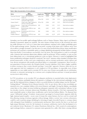
Table 3. Main characteristics of microcatheters
Main Distal inner Entry tip Crossing Delivering
Name Length (cm)
specification lumen profile profile technique
Finecross (Terumo) Single lumen, hydrophilic, 130 or 150 0.018 1.8 Fr 1.8 Fr Push without rotation
floppy tip
Corsair Pro (Asahi) Single lumen, moderated 135 or 150 0.015 1.3 Fr 2.5 Fr Counter-clockwise Rotation
hydrophilic, high support (max 10 consecutive)
Caravel (Asahi) Single lumen, highly hydrophilic 135 or 150 0.016 1.4 Fr 1.9 Fr Push without rotation
with low crossing profile
Venture (Vascular Single lumen with tip deflection 145 0.014 1.8 Fr 2.2 Fr Push without rotation
Solution) system of 90°
Crusade (Asahi) Double lumen (OTW 6.5 mm 140 0.017 2.2 Fr 2.9 Fr Push without rotation
before tip)
Twin Pass (Vascular Double lumen (OTW 11 mm 135 0.014 1.9 Fr 2.7 Fr Push without rotation
Solutions) before tip)
FineDuo (Terumo) Double lumen (OTW 6.5 mm 140 0.014 2.2 Fr 2.9 Fr Push without rotation
before the tip)
Nowadays, new low-profile rapid-exchange balloons, such as Tazuna (Terumo, Tokyo, Japan) and Ikazuchi
(Kaneka Corporation, Japan) are available. The main feature of these ballons is the extremely low entry
tip profile (between 0.015 and 0.017), lower than microcatheters, combined with a higher pushability given
by the rapid-exchange system. Therefore, the successful crossing of the lesion with a balloon rather than
microcatheter strongly increased in last few years. In cases of low-profile balloon failure, lesion modification
techniques still represent a remarkable option, as plaque debulking subsequently facilitate balloon crossing.
More specifically, if microcatheters successfully cross the lesion, the distal guidewire could be exchanged
with a rotawire and rotational atherectomy ablation could be performed. Where available, 0.9 mm excimer
laser atherectomy over a conventional guidewire is also an option. However, it must be highlighted that
lesion modification techniques should be considered “last resort” measures where standard techniques have
proved unsuccessful, as they could arise complications, such as coronary perforation and/or rupture and
even devices entrapment with possible procedural failure or catastrophic consequences. More recently a
new generation of microcatheters, the Turnpike family (Vascular Solutions, USA), show a higher capability
in lesion crossing as they contain threads enabling a “screw-like” approach. However, these devices are
quite aggressive and their employment is still relegated to CTO procedures. As expectable, most of these
equipments are now widely used in everyday PCI in every case of lesion uncrossable (due to calcification,
tortuosity or extremely narrowing) by a common semi-compliant balloon and have increased procedural
success even in daily setting.
In CTO procedures, as in everyday PCI, an adequate predilation is essential before stent deployment.
Among CTO lesions, undilatable lesions still represent a challenge for the interventional cardiologist. In the
past, the first dedicated device designed to obtain successful dilation of calcified plaque was the Flextome
TM
TM
Cutting Balloon Dilatation Device (Boston Scientific, USA). In such device, three or four microblades are
mounted over a non-compliant balloon with several diameter and length. During the dilation, microblades
create three or four plaque incisions facilitating subsequent expansion with conventional balloons. In last
two decades, however, rotational atherectomy (Rotablator, Boston Scientific, USA) have represented the
most remarkable option to obtain lesion modification and to facilitate balloon dilation. The diamond burr
causes “differential cutting” of inelastic tissues preserving integrity of normal elastic segments while the
high rotational speed (usually > 60,000 rpm) eliminate the contact between the burr and the arterial wall
thus allowing crossing of tortuous segments without damage. Seldom, excimer laser coronary atherectomy
could be used to perform plaque debulking by delivering of rapid ultraviolet B pulses to coronary lesion
with subsequent tissue breakdown by photoacoustic mechanism. More recently, the idea to use local and
high-energy lithotripsy waves for the treatment of coronary calcification lead to the development of a
new dedicated device. Shockwave IVL system (Shockwave Medical Inc., USA) consist of a semicompliant,
rapid-exchange balloon, connected to a pressure-waves generator by a cable. After lesion crossing, balloon