
Page 251 - Read Online
P. 251
Singh et al. Sternal fixation in a diabetic patient
(1) the left anterior descending artery; (2) intermediate
artery; (3) right coronary artery. The patient also had
an occluded distal circumflex artery.
At the time of angiography, the patient had an estimated
ejection fraction of about 35%. He was referred to
the care of the cardiac surgeons for consideration of
coronary artery bypass grafting (CABG).
He successfully underwent a quadruple vessel
bypass using a bilateral internal thoracic artery (BITA)
technique. This involves using both internal thoracic
arteries as a Y-graft and sequential anastomosis
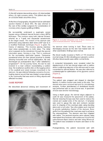
to the target vessel. The closure of his sternum was Figure 2: Computed tomography scan of thorax showing non-
union of sternum (red arrow - sternal malunion)
performed using stainless steel wires (USP size 6) in
a modified Robicsek technique [Figure 1] given his
history of diabetes. This involves placing stainless his sternum when turning in bed. There were no
steel wires parasternally on both sides. The initial discharging sinuses as the skin had healed well. All
wire is passed via the manubrium through the second other examination findings were normal.
intercostal space, forming a ring. This is continued
for the succeeding intercostal spaces. Transverse His blood results revealed a HbA1c of 110 mmol/mol
parasternal wires are then placed proximal to distally (normal range 20-42) indicating poor glycemic control.
allowing horizontal and vertical stabilization. He was All other blood results were within normal limits.
discharged on postoperative day 6 after satisfactory
progression . On his journey home, the taxi he was A computed tomography scan revealed noted the
[1]
involved in a near collision necessitating a sudden displacement of the two sternal edges with a midline
stop. He recalled an audible click on the tugging action defect confirming malunion of the sternum and he was
of the seatbelt and noted increase movement in his consented for sternal fixation. He was also referred to
sternum. He was referred following concerns of a non- a diabetologist for optimisation of his glycemic control
healing sternal wound that was treated conservatively [Figure 2].
in the community that was worst on lifting objects and
affected his sleep. Procedure
The patient was prepped and draped in standard
CASE REPORT fashion. A midline incision was made on the skin. The
sternal wires were removed and sent for microbiology
He described abnormal clicking and movement of analysis. Further debridement of devitalised tissues
was performed with no use of bone wax. A specimen
of bone was sent for microbiology.
Using a depth gauge, the sternal edges adjacent to
each rib was measured for placement of the plates.
Sternal reducing forceps was then used to reduce the
cranial and caudal ends of the sternum. A template
was used to gauge the size and contours needed
prior to shaping of the eventual plate using bending
pliers and a rod cutter. Two other plates were inserted
to provide satisfactory fixation of the sternum. Bone
grafts from the patients iliac crest was used to enhance
the osteosynthesis. The sternum was then irrigated
with saline. The pectoral muscles were sutured using
interrupted Vicryl sutures, with continuous Vicryl for the
subcutaneous layer closure and skin using Monocryl.
The patient was extubated in theatre and taken to the
wards.
Figure 1: Chest X-ray showing sternal wires Postoperatively, the patient recovered well with
Vessel Plus ¦ Volume 1 ¦ December 28, 2017 243