
Page 240 - Read Online
P. 240
Martínez et al. Cardiomyocyte energetic changes in ischemia and arrythmogenesis
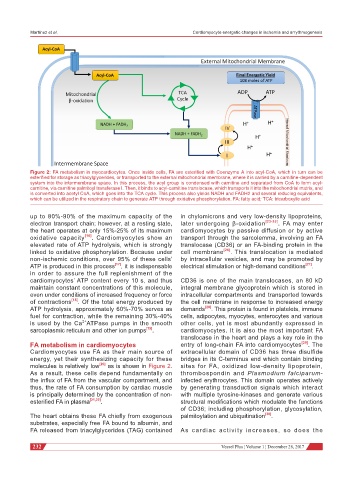
Figure 2: FA metabolism in myocardiocytes. Once inside cells, FA are esterified with Coenzyme A into acyl-CoA, which in turn can be
esterified for storage as triacylglycerides, or transported to the external mitochondrial membrane, where it is carried by a carnitine-dependent
system into the intermembrane space. In this process, the acyl group is condensed with carnitine and separated from CoA to form acyl-
carnitine, via carnitine palmitoyl transferase I. Then, it binds to acyl-carnitine translocase, which transports it into the mitochondrial matrix, and
is converted into acetyl CoA, which goes into the TCA cycle. This process also yields NADH and FADH2 and several reducing equivalents,
which can be utilized in the respiratory chain to generate ATP through oxidative phosphorylation. FA: fatty acid; TCA: tricarboxylic acid
up to 80%-90% of the maximum capacity of the in chylomicrons and very low-density lipoproteins,
electron transport chain; however, at a resting state, later undergoing β-oxidation [23-25] . FA may enter
the heart operates at only 15%-25% of its maximum cardiomyocytes by passive diffusion or by active
oxidative capacity [16] . Cardiomyocytes show an transport through the sarcolemma, involving an FA
elevated rate of ATP hydrolysis, which is strongly translocase (CD36) or an FA-binding protein in the
linked to oxidative phosphorylation. Because under cell membrane [26] . This translocation is mediated
non-ischemic conditions, over 95% of these cells’ by intracellular vesicles, and may be promoted by
ATP is produced in this process [17] , it is indispensable electrical stimulation or high-demand conditions [27] .
in order to assure the full replenishment of the
cardiomyocytes’ ATP content every 10 s, and thus CD36 is one of the main translocases, an 80 kD
maintain constant concentrations of this molecule, integral membrane glycoprotein which is stored in
even under conditions of increased frequency or force intracellular compartments and transported towards
of contractions [18] . Of the total energy produced by the cell membrane in response to increased energy
ATP hydrolysis, approximately 60%-70% serves as demands [28] . This protein is found in platelets, immune
fuel for contraction, while the remaining 30%-40% cells, adipocytes, myocytes, enterocytes and various
2+
is used by the Ca ATPase pumps in the smooth other cells, yet is most abundantly expressed in
sarcoplasmic reticulum and other ion pumps [19] . cardiomyocytes. It is also the most important FA
translocase in the heart and plays a key role in the
FA metabolism in cardiomyocytes entry of long-chain FA into cardiomyocytes [29] . The
Cardiomyocytes use FA as their main source of extracellular domain of CD36 has three disulfide
energy, yet their synthesizing capacity for these bridges in its C-terminus end which contain binding
molecules is relatively low [20] as is shown in Figure 2. sites for FA, oxidized low-density lipoprotein,
As a result, these cells depend fundamentally on thrombospondin and Plasmodium falciparum-
the influx of FA from the vascular compartment, and infected erythrocytes. This domain operates actively
thus, the rate of FA consumption by cardiac muscle by generating transduction signals which interact
is principally determined by the concentration of non- with multiple tyrosine-kinases and generate various
esterified FA in plasma [21,22] . structural modifications which modulate the functions
of CD36; including phosphorylation, glycosylation,
The heart obtains these FA chiefly from exogenous palmitoylation and ubiquitination [30] .
substrates, especially free FA bound to albumin, and
FA released from triacylglycerides (TAG) contained As cardiac activity increases, so does the
232 Vessel Plus ¦ Volume 1 ¦ December 28, 2017