Page 239 - Read Online
P. 239
Martínez et al.                                                                                                                   Cardiomyocyte energetic changes in ischemia and arrythmogenesis


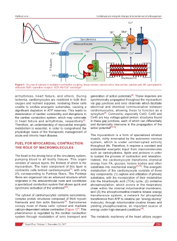
           Figure 1: The role of calcium in excitation-contraction coupling. Green arrows: calcium entry; blue arrows: calcium exit; SR: sarcoplasmic
           reticulum; RyR: ryanodine receptor; NCX: Na /Ca  exchanger
                                               2+
                                            +
           arrhythmias, heart failure, and others. During     generation of action potentials . These impulses are
                                                                                         [8]
           ischemia, cardiomyocytes are restricted in both their   synchronically propagated throughout the myocardium
           oxygen and nutrient supplies, rendering these cells   via gap junctions and ionic channels which facilitate
           unable to oxidize energetic substrates, causing a   electrical and chemical communication between
           significant depletion in ATP reserves. This leads to   cardiomyocytes, allowing these to function as a
                                                                       [9]
           deterioration of cardiac contractility and disruptions in   syncytium . Connexins, especially Cx43, Cx40 and
           the cardiac conduction system, which may culminate   Cx45 are key voltage-gated proteic structures found
                                                         [2]
           in heart failure and arrhythmias, respectively .   in these gap junctions, each of which can differentially
           Therefore, an understanding of myocardial energetic   and dynamically intervene in the propagation of the
           metabolism is essential, in order to comprehend the   action potential [10-12] .
           physiologic basis of the therapeutic management of
           acute and chronic heart disease.                   The myocardium is a form of specialized striated
                                                              muscle, richly innervated by the autonomic nervous
           FUEL FOR MYOCARDIAL CONTRACTION:                   system, which is under uninterrupted activity
           THE ROLE OF MACROMOLECULES                         throughout life. Therefore, it requires a constant and
                                                              substantial energetic input from macromolecules
                                                              such as carbohydrates, lipids and proteins in order
           The heart is the driving force of the circulatory system,   to sustain the process of contraction and relaxation.
           pumping blood to all bodily tissues. This organ    Indeed,  the  cardiomyocyte  transforms  chemical
           consists of various layers, the thickest of which is the   energy from FA, glucose, ketone bodies and other
           myocardium. The main component of this layer is    substrates into mechanical energy [13,14] . The energetic
           contractile cells termed cardiomyocytes , with only   metabolism of the cardiomyocyte consists of three
                                                [3]
           2% corresponding to Purkinje fibers. The Purkinje   key components: (1) capture and utilization of primary
           fibers are organized into an arborized structure which   substrates, with the incorporation of their metabolites
           originates in the atrioventricular node and constitutes   into the tricarboxylic acid (TCA) cycle; (2) oxidative
           a specialized conduction system that allows quick and   phosphorylation, which occurs in the respiratory
           synchronic activation of the ventricles [4,5] .    chain within the internal mitochondrial membrane;
                                                              and (3) the phosphocreatine-creatine kinase energy
           The cytosol of cardiomyocytes contains sarcomeres,   transference system, a network for phosphate
           complex proteic structures composed of thick myosin   transference from ATP to creatine (an “energy-storing”
                                            [6]
           filaments and thin actin filaments . Sarcomeres    molecule), through mitochondrial creatine kinase and
           occupy most of these cells’ cytosol and mediate    yielding phosphocreatine, an important source of
           calcium-dependent cellular contraction [Figure 1] . This   energy under high-demand conditions [15] .
                                                     [7]
           phenomenon is regulated by the cardiac conduction
           system through modulation of ionic transport and   The metabolic machinery of the heart utilizes oxygen

                           Vessel Plus ¦ Volume 1 ¦ December 28, 2017                                     231

