
Page 168 - Read Online
P. 168
Bertellotti et al. Vertebral artery transection
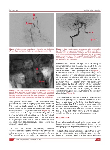
Figure 1: Vertebral artery surgically controlled prior to embolization Figure 2: Right vertebral artery angiography after embolization
(A) and digital subtraction shadow of surgically applied clips (B) by coils of the very proximal part of left vertebral artery (arrow
showing coils, B). Retrograde opacity of the distal left vertebral
artery shows persistent extravasation of contrast with an extradural
pseudoaneurysm above the surgically placed clips which had
controlled all bleeding operatively (A) secondary to vessel
transection from the stab wound
micro-catheter through the right vertebral artery in
retrograde fashion into the very distal part of the left
vertebral artery with navigation of the catheter tip
above the level of the surgically placed clips [Figure 2].
Embolization in this location was performed again by
lumen occlusion with coils with meticulous preservation
of the anterior spinal artery, which had its origin from
the distal left vertebral artery. The territory of the left
posterior inferior cerebellar artery was satisfactory
collateralized by the left anterior inferior cerebellar
artery. Thus, this endovascular approach obtained
complete proximal and distal trapping of the left
Figure 3: The micro catheter was placed in retrograde fashion in vertebral artery pseudoaneurysm above the surgically
the left distal vertebral artery immediately above the extradural
pseudoaneurysm during deployment. Endovascular approach placed clips [Figure 3].
obtained complete proximal (A) and distal (B) trapping of the extra
dural left vertebral artery pseudoaneurysm above the surgical clips The patient was transferred to the ICU, extubated on
the second post-operative day and transferred to the
Angiographic visualization of the vasculature was floor. He was observed for 2 days and discharged on
performed by catheter angiography, which revealed post-operative day 4. No problems were noted with
that complete acute occlusion of the left vertebral balance when ambulating. He had an uneventful
artery at the C1-C2 level was successfully achieved recovery. He was evaluated in the Trauma Clinic during
during the second surgical intervention [Figure 1]. The his 7, 14, 30 and 60-day follow-up with no sequelae
catheterization of the right vertebral artery showed noted from this rare and complex vascular injury.
normal perfusion with opacification of the very distal
segment of the left vertebral artery. The late phase DISCUSSION
of angiography depicted a pattern consistent with
an extra-dural pseudoaneurysm noted above the Penetrating vertebral artery injuries are rare and their
surgically placed clips. injuries were previously missed prior to the routine use
of angiography in diagnosing penetrating neck injuries.
The first step during angiography included
endovascular embolization by coils of the left vertebral This patient specifically, sustained a penetrating injury
artery proximal to the visualized surgical occlusion. to the vertebral artery and had hard signs of vascular
The second stage proceeded by navigation of the injury with profuse bleeding at the scene and upon
Vessel Plus ¦ Volume 1 ¦ September 26, 2017 161