
Page 159 - Read Online
P. 159
Shaikhrezai et al. Late presenting valve endocarditis
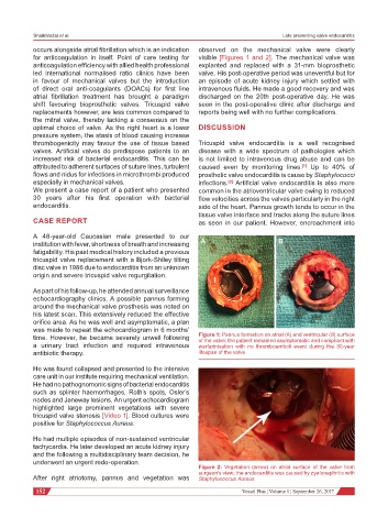
occurs alongside atrial fibrillation which is an indication observed on the mechanical valve were clearly
for anticoagulation in itself. Point of care testing for visible [Figures 1 and 2]. The mechanical valve was
anticoagulation efficiency with allied health professional explanted and replaced with a 31-mm bioprosthetic
led international normalised ratio clinics have been valve. His post-operative period was uneventful but for
in favour of mechanical valves but the introduction an episode of acute kidney injury which settled with
of direct oral anti-coagulants (DOACs) for first line intravenous fluids. He made a good recovery and was
atrial fibrillation treatment has brought a paradigm discharged on the 20th post-operative day. He was
shift favouring bioprosthetic valves. Tricuspid valve seen in the post-operative clinic after discharge and
replacements however, are less common compared to reports being well with no further complications.
the mitral valve, thereby lacking a consensus on the
optimal choice of valve. As the right heart is a lower DISCUSSION
pressure system, the stasis of blood causing increase
thrombogenicity may favour the use of tissue based Tricuspid valve endocarditis is a well recognised
valves. Artificial valves do predispose patients to an disease with a wide spectrum of pathologies which
increased risk of bacterial endocarditis. This can be is not limited to intravenous drug abuse and can be
attributed to adherent surfaces of suture lines, turbulent caused even by monitoring lines. Up to 40% of
[1]
flows and nidus for infections in microthrombi produced prosthetic valve endocarditis is cause by Staphylococci
especially in mechanical valves. infections. Artificial valve endocarditis is also more
[2]
We present a case report of a patient who presented common in the atrioventricular valve owing to reduced
30 years after his first operation with bacterial flow velocities across the valves particularly in the right
endocarditis. side of the heart. Pannus growth tends to occur in the
tissue valve interface and tracks along the suture lines
CASE REPORT as seen in our patient. However, encroachment into
A 48-year-old Caucasian male presented to our
institution with fever, shortness of breath and increasing A B
fatigability. His past medical history included a previous
tricuspid valve replacement with a Bjork-Shiley tilting
disc valve in 1986 due to endocarditis from an unknown
origin and severe tricuspid valve regurgitation.
As part of his follow-up, he attended annual surveillance
echocardiography clinics. A possible pannus forming
around the mechanical valve prosthesis was noted on
his latest scan. This extensively reduced the effective
orifice area. As he was well and asymptomatic, a plan
was made to repeat the echocardiogram in 6 months’
time. However, he became severely unwell following Figure 1: Pannus formation on atrial (A) and ventricular (B) surface
of the valve; the patient remained asymptomatic and compliant with
a urinary tract infection and required intravenous warfarinisation with no thromboemboli event during the 30-year
antibiotic therapy. lifespan of the valve
He was found collapsed and presented to the intensive
care unit in our institute requiring mechanical ventilation.
He had no pathognomonic signs of bacterial endocarditis
such as splinter haemorrhages, Roth’s spots, Osler’s
nodes and Janeway lesions. An urgent echocardiogram
highlighted large prominent vegetations with severe
tricuspid valve stenosis [Video 1]. Blood cultures were
positive for Staphylococcus Aureus.
He had multiple episodes of non-sustained ventricular
tachycardia. He later developed an acute kidney injury
and the following a multidisciplinary team decision, he
underwent an urgent redo-operation.
Figure 2: Vegetation (arrow) on atrial surface of the valve from
surgeon’s view; the endocarditis was caused by pyelonephritis with
After right atriotomy, pannus and vegetation was Staphylococcus Aureus
152 Vessel Plus ¦ Volume 1 ¦ September 26, 2017