
Page 66 - Read Online
P. 66
Page 4 of 11 Mattana et al. Vessel Plus 2022;6:13 https://dx.doi.org/10.20517/2574-1209.2021.87
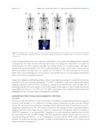
Figure 3. Technetium-99m 3,3-diphosphono-1,2-propanodicarboxylic acid bone scan, 3 h acquisition: (A) visual score 0; (B) visual
score 1 and corresponding single photon emission computed tomography/computed tomography image; (C) visual score 2; and (D)
visual score 3.
Despite the high diagnostic accuracy reported in the literature, CA remains underdiagnosed and sometimes
misdiagnosed. The most common and important cause of misdiagnosis is the failure to exclude AL
cardiomyopathy (AL-CM) in patients with high tracer uptake (Grade 2 or 3 on planar images). Although in
the literature myocardial uptake in AL-CM is usually described as Grade 1 or less, larger cohorts reported
that 22% of patients showing Grade 2 or 3 uptakes on Tc-DPD or Tc-PYP scans have AL-CM on the
99m
99m
EMB. Other cases of misdiagnosis can be related to some pitfalls due to a wrong imaging interpretation
and/or an uncorrected acquisition protocol.
Despite the emphasis in published guidelines, failure or incomplete screening for AL amyloidosis has been
reported in up to 24% of patients undergoing Tc-PYP ; in addition, many centers only perform planar
[20]
99m
imaging acquisition (without SPECT imaging to confirm myocardial uptake), with the high risk of
misinterpreting the blood pool uptake as myocardial uptake. In this paper, we aim to briefly describe the
main differences between Tc-PYP and Tc-DPD, analyzing the data available in the literature and
99m
99m
highlighting the most frequent causes of misdiagnosis and pitfalls.
ACQUISITION PROTOCOLS AND DIAGNOSTIC CRITERIA
99m
Tc-PYP
This tracer is the only used tracer in the United States. Nevertheless, a standardized and shared acquisition
protocol is still lacking so it is difficult to correctly compare and analyze the data from the different
[21]
[22]
studies. The American Society of Nuclear Cardiology (ASNC) released the latest Practice Points in 2019 ,
99m
suggesting to standardize the Tc-PYP acquisition protocol. The writing committee recommended planar
imaging followed by the single-photon emission computerized tomography (SPECT) imaging or
SPECT/CT imaging at 1 h; an optional SPECT imaging at 3 h can be used when a high blood pool activity
has been observed in the 1 h SPECT images. The semi-quantitative analysis criteria reckon on a heart to
contralateral lung ratio (H/CL) > 1.5 on 1 h imaging to accurately identify ATTR cardiac amyloidosis if
systemic AL amyloidosis has been excluded. A similar recommendation was reported in a multi-societal
expert consensus on imaging CA .
[23]