
Page 68 - Read Online
P. 68
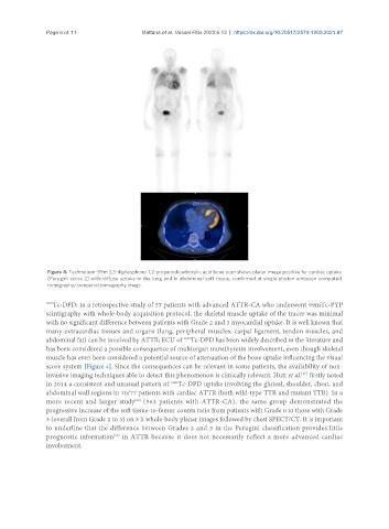
Page 6 of 11                 Mattana et al. Vessel Plus 2022;6:13  https://dx.doi.org/10.20517/2574-1209.2021.87


                Figure 4. Technetium-99m 3,3-diphosphono-1,2-propanodicarboxylic acid bone scan shows planar image positive for cardiac uptake
                (Perugini score 2) with diffuse uptake in the lung and in abdominal soft tissue, confirmed at single photon emission computed
                tomography/computed tomography image.

               99m Tc-DPD: in a retrospective study of 57 patients with advanced ATTR-CA who underwent 99mTc-PYP
               scintigraphy with whole-body acquisition protocol, the skeletal muscle uptake of the tracer was minimal
               with no significant difference between patients with Grade 2 and 3 myocardial uptake. It is well known that
               many extracardiac tissues and organs (lung, peripheral muscles, carpal ligament, tendon muscles, and
               abdominal fat) can be involved by ATTR; ECU of  Tc-DPD has been widely described in the literature and
                                                         99m
               has been considered a possible consequence of multiorgan transthyretin involvement, even though skeletal
               muscle has even been considered a potential source of attenuation of the bone uptake influencing the visual
               score system [Figure 4]. Since the consequences can be relevant in some patients, the availability of non-
               invasive imaging techniques able to detect this phenomenon is clinically relevant. Hutt et al.  firstly noted
                                                                                             [27]
               in 2014 a consistent and unusual pattern of  Tc-DPD uptake involving the gluteal, shoulder, chest, and
                                                     99m
               abdominal wall regions in 70/77 patients with cardiac ATTR (both wild-type TTR and mutant TTR). In a
                                          [28]
               more recent and larger study  (563 patients with ATTR-CA), the same group demonstrated the
               progressive increase of the soft tissue-to-femur counts ratio from patients with Grade 0 to those with Grade
               3 (overall from Grade 2 to 3) on 3 h whole-body planar images followed by chest SPECT/CT. It is important
               to underline that the difference between Grades 2 and 3 in the Perugini classification provides little
               prognostic information  in ATTR because it does not necessarily reflect a more advanced cardiac
                                    [28]
               involvement.