
Page 70 - Read Online
P. 70
Page 8 of 11 Mattana et al. Vessel Plus 2022;6:13 https://dx.doi.org/10.20517/2574-1209.2021.87
Figure 6. Case of a 60-year-old male with echocardiography consistent to hypertrophic cardiomyopathy, preserved ejection fraction.
Pericardial effusion. Technetium-99m 3,3-diphosphono-1,2-propanodicarboxylic acid bone scan performed for clinical suspicion of
cardiac amyloidosis revealed mild uptake in heart area, but single photon emission computed tomography/computed tomography
demonstrated that it is due to blood pool activity: visual score 0.
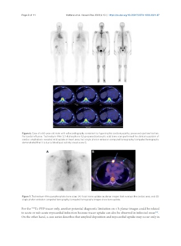
Figure 7. Technetium-99m pyrophosphate bone scan: (A) focal tracer uptake on planar images that overlaps the cardiac area; and (B)
single photon emission computed tomography/computed tomography images show bone uptake.
99m
For the Tc-PYP tracer only, another potential diagnostic limitation on 1 h planar images could be related
[34]
to acute or sub-acute myocardial infarction because tracer uptake can also be observed in infarcted areas .
On the other hand, a case series describes that amyloid deposition and myocardial uptake may occur only in