


Page 68 - Read Online
P. 68
Cepas-Guillen et al. Vessel Plus 2021;5:26  https://dx.doi.org/10.20517/2574-1209.2020.79  Page 5 of 12

               Table 1. Anatomical indications/restrictions for percutaneous mitral “edge-to-edge” therapies
                Optimal anatomy          Suboptimal anatomy            Unsuitable anatomy
                Central pathology in segment 2  Pathology in segment 1 or 3  Perforated MV leaflet or cleft
                No leaflet calcification  Mild calcification outside of the grip-zone   Severe calcification in the grip-zone
                                         Ring calcification
                                         Post annuloplasty
                         2                        2                             2
                MVOA > 4 cm              MVOA > 3 cm                   MVOA < 3 cm , MGP ≥ 5 mmHg
                Mobile length of PML ≥ 10 mm  Mobile length of PML 7-10 mm  Mobile length of PML < 7 mm
                Coaptation Depth < 11 mm  Coaptation Depth ≥ 11 mm     Rheumatic leaflet thickening
                                                                       Restriction in systole and diastole (Carpentier IIIa)
                Normal leaflet strength and mobility  Leaflet restriction in systole   Barlow’s syndrome
                                         (Carpentier IIIb)
                Flail width < 15 mm      Flail width > 15 mm
                Flail gap < 10 mm

               MV: Mitral valve; MVOA: mitral valve orifice area; MPG: mean pressure gradient.


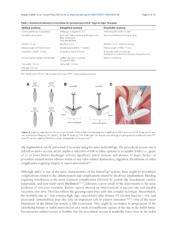
                Figure 2. Edge-to-edge device: (A) four size options of MitraClip® G4 including two length sizes [NT (short-arm)/XT (long-arm)] and
                two width sizes [Regular/W (wide)]; (B) the W family is 1.5× wider than the regular clip offering a larger grasping width with both NT
                                                             [7]
                and XT options; and (C) PASCAL system. Adapted from Pozzoli et al. .
               clip implantation can be performed if necessary using the same methodology. The procedural success rate is
               defined as device success, which implies a reduction of MR to either optimal or acceptable levels ( i.e., grade
               of 2+ or lower before discharge) without significant mitral stenosis and absence of major device- or
               procedure-related serious adverse events or any valve-related dysfunction, migration, thrombosis, or other
                                                            [28]
               complication requiring surgery or repeat intervention .
               Although safety is one of the main characteristics of the MitraClip® system, there might be procedural
               complications related to the catheterization and complications related to the device implantation. Bleeding
               requiring transfusion is the most common complication followed by partial clip detachment, cardiac
               tamponade, and new-onset atrial fibrillation [2,15,19] . Likewise, a poor result of the intervention is the main
               predictor of one-year mortality. Recent reports showed an improvement in success rate and durable
               outcomes over time. This fact reflects the growing experience with this complex technique. Nevertheless,
               the mortality rate at 1 year remains high. Age, concomitant valve disease, LV ejection fraction < 30%, and
               associated comorbidities may also play an important role in patient outcomes [18,21,29] . One of the main
               limitations of the MitraClip system is MR recurrence. This might be secondary to progression of the
               underlying disease or leaflet insertion loss as a result of insufficient capture of the clip in the leaflet tissue.
               Percutaneous reintervention is feasible, but the procedural success is markedly lower than in the index

