


Page 66 - Read Online
P. 66
Cepas-Guillen et al. Vessel Plus 2021;5:26  https://dx.doi.org/10.20517/2574-1209.2020.79  Page 3 of 12


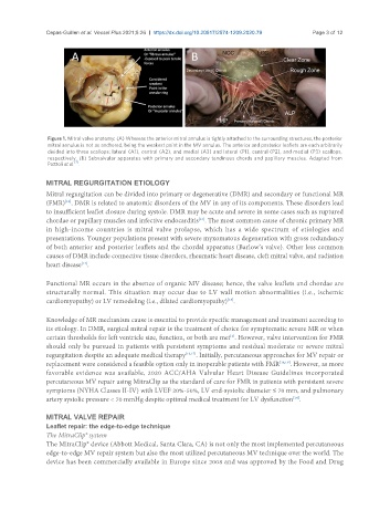
                Figure 1. Mitral valve anatomy: (A) Whereas the anterior mitral annulus is tightly attached to the surrounding structures, the posterior
                mitral annulus is not as anchored, being the weakest point in the MV annulus. The anterior and posterior leaflets are each arbitrarily
                divided into three scallops: lateral (A1), central (A2), and medial (A3) and lateral (P1), central (P2), and medial (P3) scallops,
                respectively. (B) Subvalvular apparatus with primary and secondary tendinous chords and papillary muscles. Adapted from
                        [7]
                Pozzoli et al. .
               MITRAL REGURGITATION ETIOLOGY
               Mitral regurgitation can be divided into primary or degenerative (DMR) and secondary or functional MR
               (FMR) . DMR is related to anatomic disorders of the MV in any of its components. These disorders lead
                     [12]
               to insufficient leaflet closure during systole. DMR may be acute and severe in some cases such as ruptured
               chordae or papillary muscles and infective endocarditis . The most common cause of chronic primary MR
                                                              [13]
               in high-income countries is mitral valve prolapse, which has a wide spectrum of etiologies and
               presentations. Younger populations present with severe myxomatous degeneration with gross redundancy
               of both anterior and posterior leaflets and the chordal apparatus (Barlow’s valve). Other less common
               causes of DMR include connective tissue disorders, rheumatic heart disease, cleft mitral valve, and radiation
               heart disease .
                          [14]
               Functional MR occurs in the absence of organic MV disease; hence, the valve leaflets and chordae are
               structurally normal. This situation may occur due to LV wall motion abnormalities (i.e., ischemic
               cardiomyopathy) or LV remodeling (i.e., dilated cardiomyopathy) .
                                                                      [15]

               Knowledge of MR mechanism cause is essential to provide specific management and treatment according to
               its etiology. In DMR, surgical mitral repair is the treatment of choice for symptomatic severe MR or when
               certain thresholds for left ventricle size, function, or both are met . However, valve intervention for FMR
                                                                       [2]
               should only be pursued in patients with persistent symptoms and residual moderate or severe mitral
               regurgitation despite an adequate medical therapy [16,17] . Initially, percutaneous approaches for MV repair or
               replacement were considered a feasible option only in inoperable patients with FMR [18,19] . However, as more
               favorable evidence was available, 2020 ACC/AHA Valvular Heart Disease Guidelines incorporated
               percutaneous MV repair using MitraClip as the standard of care for FMR in patients with persistent severe
               symptoms (NYHA Classes II-IV) with LVEF 20%-50%, LV end-systolic diameter ≤ 70 mm, and pulmonary
               artery systolic pressure < 70 mmHg despite optimal medical treatment for LV dysfunction .
                                                                                          [14]

               MITRAL VALVE REPAIR
               Leaflet repair: the edge-to-edge technique
               The MitraClip® system
               The MitraClip® device (Abbott Medical, Santa Clara, CA) is not only the most implemented percutaneous
               edge-to-edge MV repair system but also the most utilized percutaneous MV technique over the world. The
               device has been commercially available in Europe since 2008 and was approved by the Food and Drug

