


Page 72 - Read Online
P. 72
Cepas-Guillen et al. Vessel Plus 2021;5:26  https://dx.doi.org/10.20517/2574-1209.2020.79  Page 9 of 12


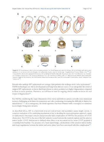
                Figure 4. (1) Percutaneous mitral valve anchoring systems: (a) Atrial flange and ventricle tethers; (b) atrial flange and native valve
                anchors; (c) atrial and ventricle flanges; (d) subannular hooks; and (e) atrial cage. Adapted from Preston-Maher  et al. [46] . (2)
                Transcatheter mitral valve replacement devices. (A) CardiAQ/EVOQUE (Edwards Lifesciences Inc); (B) Tiara (Neovasc Inc, Canada);
                (C) Tendyne (Abbott Inc); (D) Intrepid (Medtronic Inc); (E) Caisson (LivaNova, UK); (F) HighLife Bioprosthesis and Subannular
                Implant (HighLife SAS, France); (G) SAPIEN M3 (Edwards Lifesciences Inc); and (H) Cardiovalve (Cardiovalve, Israel). Adapted from
                Testa et al. [47] .

               Patients who undergo MV replacement are younger than patients who undergo aortic valve replacement .
                                                                                                        [1]
               TMVR technologies are still in development and long-term data are scarce. If we extrapolate the result of
               surgical MV replacement, we know that bioprotheses in mitral position have higher degeneration compared
               to those in aortic position. Therefore, we will have to wait for ongoing clinical studies to know the real
               impact of degeneration .
                                   [47]

               For TMVR, considering the larger dimensions of the valves and delivery system currently used, transseptal
               access is challenging as its limits the maneuvers and valve positioning, increasing the difficulty to deploy the
               mitral device [47,48] . As a consequence, the initial experience has been obtained with a transapical or transatrial
               access despite being more invasive.


               As described above, MV is a functional structure and primary and secondary causes might coexist. A
               complete evaluation of the underlying mechanism is key to deciding the most appropriate approach: repair
               or replacement. One major concern and potentially fatal complication of TMVR is the presence of LVOT
               obstruction. The LVOT is the area of the left ventricle located between the ventricle septum and the anterior
               mitral leaflet. LVOT obstruction is defined when there is an increment in the mean LVOT gradient ≥
               10 mmHg from baseline. The presence of a basal septal budge, calcifications of the anterior mitral leaflet,
               and sharp angulations between the mitral and aortic plane have been associated with this complication .
                                                                                                       [49]

