
Page 22 - Read Online
P. 22
Page 8 of 14 Ricci et al. Vessel Plus 2021;5:31 https://dx.doi.org/10.20517/2574-1209.2021.28
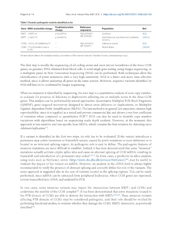
Table 1. Founder pathogenic variants identified so far
Predicted protein Reference
Gene DNA nucleotide change Population Ref.
change sequences
KRIT1 c.987C>A p.Cys329Ter NM_194456.1 Sardinian [62]
NP_919438.1
KRIT1 c.1363C>T p.Gln455Ter North Mexican and American Southwest [58,59]
ancestry
CCM2 c.30+5_30+6delGCinsTT -- NM_031443.3 Ashkenazi Jewish [63]
NP_113631.1
CCM2 77.6-kb deletion (exons -- United States [38,60]
2–10)
Nomenclature follows the standard naming conventions of the Human Genomic Variation Society (varnomen.hgvs.org).
The first step is usually the sequencing of all coding exons and exon-intron boundaries of the three CCM
genes, on genomic DNA obtained from blood cells. A serial single gene testing, using Sanger sequencing, or
a multigene panel in Next Generation Sequencing (NGS) can be performed. Both techniques allow the
identification of point mutations with a very high sensitivity. NGS is a faster and more time-effective
method, since it allows analyzing all genes in the same session. However, sequence variants identified by
NGS still have to be confirmed by Sanger sequencing.
When no mutation is identified by sequencing, the next step is a quantitative analysis of exon copy number,
to evaluate the presence of deletions or duplications affecting one or multiple exons in the three CCM
genes. This analysis can be performed by several approaches: Quantitative Multiplex PCR Short Fragments
(QMPSF), gene-targeted microarray designed to detect exon deletions or duplications, or Multiplex
Ligation-dependent Probe Amplification (MLPA). The last method is in general less expensive; ensures high
reproducibility, since it is supplied as a tested and proven commercial kit; and shows a very low coefficient
[70]
of variation when compared to quantitative PCR . NGS can also be used to identify copy number
variations with algorithms based on sequencing reads depth analysis. However, at the moment, this
approach is less sensitive and less specific than MLPA, which remains the best solution for detecting exon
deletion/duplication .
[71]
If a variant is identified in the first two steps, its role has to be evaluated. If the variant introduces a
premature stop codon (nonsense or frameshift variant, caused by point mutations or exon deletions) or is
located in an invariant splicing region, its pathogenic role is easy to define. The pathogenic features of
missense mutations are more difficult to establish. Indeed, it has been demonstrated that some “missense”
mutations actually activate cryptic splice sites and cause an aberrant splicing of CCM mRNA, resulting in
frameshift and introduction of a premature stop codon [1,72,73] . In these cases, a predictive in silico analysis,
using tools such as NetGene2 server (http://www.cbs.dtu.dk/services/NetGene2) , may be useful to
[74]
evaluate the impact of the variant on mRNA. However, an analysis at the cDNA level is always highly
recommended to verify the presence of aberrant splicing and correctly define the role of the variants. The
same approach is suggested also in the case of variants located in the splicing regions. This can be easily
performed, since mRNA can be extracted from peripheral leukocytes, where CCM genes are expressed,
reverse transcribed into cDNA, and analyzed by PCR.
In rare cases, some missense variants may impair the interactions between KRIT1 and CCM2 and
undermine the stability of the CCM complex . It has been demonstrated that some mutations located in
[39]
the PTB domain of CCM2 are able to destroy the interaction with KRIT1 [38,40,41] . Thus, missense variants
affecting PTB domain of CCM2 may be considered pathogenic, and their role should be verified by
performing functional studies, to evaluate whether they damage the CCM2-KRIT1 interaction, as previously
described .
[39]