
Page 24 - Read Online
P. 24
Page 10 of 14 Ricci et al. Vessel Plus 2021;5:31 https://dx.doi.org/10.20517/2574-1209.2021.28
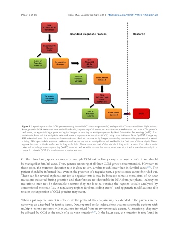
Figure 7. Stepwise protocol of CCM gene screening in familial CCM cases (probands) and sporadic CCM cases with multiple lesions.
After genomic DNA extraction from white blood cells, sequencing of all exons and intron-exon boundaries of the three CCM genes is
performed, using serial single gene testing by Sanger sequencing or multigene panels by Next Generation Sequencing (NGS). If no
mutation is detected, the analysis is extended to exon copy number variations (CNV) using quantitative MLPA or QMPSF. If negative,
RNA extracted from blood leukocytes is reverse transcribed and sequenced by Sanger sequencing to evaluate the presence of aberrant
splicing. This approach is also used in the case of variants of uncertain significance identified in the first step of the analysis. These
approaches are routinely performed in diagnostic labs. These steps are part of the standard diagnostic process. If no alteration is
detected, whole genome sequencing (WGS) may be performed to assess the presence of rare structural anomalies (usually in a
research context). CCM: Cerebral cavernous malformations.
On the other hand, sporadic cases with multiple CCM lesions likely carry a pathogenic variant and should
be managed as familial cases. Thus, genetic screening of all three CCM genes is recommended. However, in
these cases, the mutation detection rate is close to 60%, a value much lower than in familial cases [13,78] . The
patient should be informed that, even in the presence of a negative test, a genetic cause cannot be ruled out.
There can be several explanations for a negative test: it may be because somatic mosaicism of de novo
mutations occurred during gestation and therefore are not detectable in DNA from peripheral leukocytes;
mutations may not be detectable because they are located outside the regions usually analyzed by
conventional methods (i.e., in regulatory regions far from coding exons); and epigenetic modifications able
to alter the expression of CCM proteins may occur.
When a pathogenic variant is detected in the proband, the analysis may be extended to the parents, in the
same way as described for familial cases. Data reported so far indeed show that most sporadic patients with
multiple lesions are cases with mutations inherited from an asymptomatic parent. Alternatively, they may
be affected by CCM as the result of a de novo mutation [1,77] . In the latter case, the mutation is not found in