


Page 51 - Read Online
P. 51
Trahanas et al. Vessel Plus 2022;6:49  https://dx.doi.org/10.20517/2574-1209.2021.125  Page 11 of 16


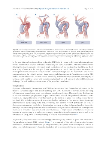
                Figure 6. Artist drawings of type I and II hybrid arch repairs (HAR) for chronic dissection. Type I: HAR involves debranching of the aortic
                arch vessels using a multi-branched Dacron graft sewn to either the native ascending aorta or, as shown in the drawing, a previously
                Dacron-replaced ascending aorta. This latter scenario is most commonly utilized in the setting of residual type B dissection after prior
                type A repair. Type II: HAR involves the replacement of the ascending aorta and, most commonly, a portion of the arch (to create
                proximal landing zone for the endografts) with distal anastomosis frequently in zones 1 or 2, along with debranching of the arch vessels.

               In the near future, physician-modified endografts (PMEGs) and visceral multi-branched endografts may
               become an alternative to hybrid abdominal debranching and TEVAR in unfit CTBAD patients with disease
               affecting the visceral segment; a prior small, single-institution study has confirmed the feasibility and short-
               term safety of PMEGs in the setting of chronic post-dissection TAAA . Briefly, at our institution, the
                                                                             [45]
               PMEG procedure involves the modification of an off-the-shelf endograft via the creation of 4 fenestrations
               corresponding to the patient’s anatomy based upon detailed measurements from the preoperative CTA
               study . Patient selection for PMEG is critical. Specifically, suitable anatomy is paramount, as attempting to
                    [46]
               navigate a stiff small true lumen with branches originating from both lumens is a formidable technical
               challenge. The mid- and long-term outcomes of the procedure are unknown.


               Complications
               Open and endovascular interventions for CTBAD are not without risk. Potential complications are like
               those of any aortic surgery and include inducing new aortic dissection or rupture, stroke, bleeding,
               infection, acute kidney injury, bowel ischemia, and wound complications. The complication that is unique
               to aortic intervention is paraplegia due to spinal cord ischemia. The risk for spinal cord ischemia increases
               based on the length of the aorta covered/replaced, and the number of spinal cord feeding vessels that are
               sacrificed. This includes the vertebral, intercostal, lumbar, and internal iliac arteries. Neurophysiologic
               intraoperative  monitoring  with  somatosensory  and  motor  evoked  potentials,  as  well  as
               electroencephalography, can help to detect spinal cord and cerebral ischemia. Serial postoperative
               neurologic exams are also paramount to detect early evidence of proximal muscle weakness. Intra-operative
               measures to prevent or treat spinal cord ischemia include re-implantation of lumbar and intercostal
               branches in open surgery, or in endovascular surgery revascularization of any covered arteries such as the
               left subclavian artery, which is the major supply of collateral flow to the spinal cord [3,26,27] .

               A systematic perioperative approach should be applied to manage any evidence of spinal cord compromise.
               The paraplegia treatment COPS protocol [Figure 7] is an example of this: with a focus on blood pressure
               stabilization, cerebrospinal fluid drainage, oxygen delivery, and patient status leading to an increase in
               spinal cord perfusion pressure . Such standardization allows close monitoring and more rapid treatment
                                         [47]

