


Page 49 - Read Online
P. 49
Sciahbasi et al. Vessel Plus 2023;7:19  https://dx.doi.org/10.20517/2574-1209.2023.22  Page 9 of 12


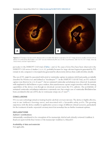
                Figure 3. OCT image of severe calcific stenosis before and after IVL balloon expansion. (A) OCT image of severe calcific stenosis with an
                almost circumferential distribution over 260° (as also indicated by the arcs on the circumference). (B) Post IVL OCT image, showing
                intracoronary calcium fragmentation.

               particular in the DISRUPT CAD trials [Tables 1 and 2]. No cases of slow flow have been observed in the
               DISRUPT CAD series of studies (12,25-27), probably because the large calcium fragments generated by IVL
               remain in situ compared to microparticles generated by atherectomy devices that could embolize distally.


               The use of IVL might be associated with atrial or ventricular capture in patients with bradycardia, as initially
               described by Wilson et al. and defined as “shocktopics” . In the DISRUPT CAD III Trial, an IVL-induced
                                                              [51]
                                              [13]
               capture was observed in 41% of cases , but no sustained ventricular arrhythmias were observed. In patients
               with implantable defibrillators or pace-makers, this asynchronous capture might interfere with the sensing
               capabilities of the device even though no electrical current leaves the IVL catheter. The probability of
               sustained ventricular arrhythmias induction is extremely rare, but a single case of ventricular fibrillation has
               been correlated to the use of IVL due to ventricular ectopy on a T wave .
                                                                           [52]

               CONCLUSIONS
               IVL is a new technology aimed at treating heavily calcified coronary stenosis. The device is highly effective,
               easy to use (without a learning curve), and associated with a favourable safety profile. The growing
               experience with the device enables its application across a range of different clinical scenarios, particularly
               for the treatment of under-expanded coronary stent that nowadays has no further treatment options.


               DECLARATIONS
               Authors’ contributions
               Substantially contributed to the conception of the manuscript, drafted and critically reviewed: Sciahbasi A
               Read and approved the final version of the manuscript: Sciahbasi A, Minardi S

               Availability of data and materials
               Not applicable.

