


Page 47 - Read Online
P. 47
Sciahbasi et al. Vessel Plus 2023;7:19  https://dx.doi.org/10.20517/2574-1209.2023.22  Page 7 of 12


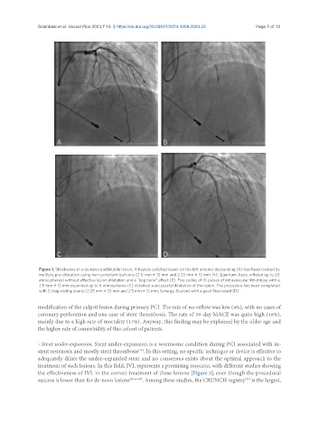
                Figure 1. Shockwave in a de-novo undilatable lesion. A heavily calcified lesion on the left anterior descending (A) has been treated by
                multiple pre-dilatation using non-compliant balloons (2.0 mm × 15 mm and 2.25 mm × 15 mm, NC Quantum Apex, inflated up to 20
                atmospheres) without effective lesion dilatation and a “dog bone” effect (B). Five cycles of 10 pulses of intravascular lithotripsy with a
                2.5 mm × 12 mm expanded up to 6 atmospheres (C) obtained a successful dilatation of the lesion. The procedure has been completed
                with 2 drug eluting stents (2.25 mm × 32 mm and 2.5 mm × 12 mm, Synergy, Boston) with a good final result (D).


               modification of the culprit lesion during primary PCI. The rate of no-reflow was low (4%), with no cases of
               coronary perforation and one case of stent thrombosis. The rate of 30-day MACE was quite high (18%),
               mainly due to a high rate of mortality (17%). Anyway, this finding may be explained by the older age and
               the higher rate of comorbidity of this cohort of patients.

               - Stent under-expansion. Stent under-expansion is a worrisome condition during PCI associated with in-
               stent restenosis and mostly stent thrombosis . In this setting, no specific technique or device is effective to
                                                     [42]
               adequately dilate the under-expanded stent and no consensus exists about the optimal approach to the
               treatment of such lesions. In this field, IVL represents a promising resource, with different studies showing
               the effectiveness of IVL in the correct treatment of these lesions [Figure 3], even though the procedural
                                                                                            [46]
               success is lower than for de-novo lesions [28,43-45] . Among these studies, the CRUNCH registry  is the largest,

