


Page 22 - Read Online
P. 22
Page 4 of 22                   Andò et al. Vessel Plus 2023;7:15  https://dx.doi.org/10.20517/2574-1209.2023.26


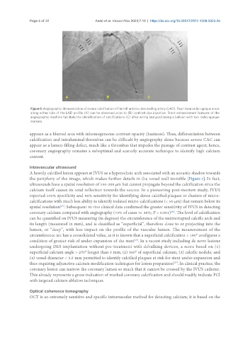
                Figure 1. Angiographic demonstration of severe calcification of the left anterior descending artery (LAD). Two linear radio-opaque areas
                along either side of the LAD profile (A) can be observed prior to (B) contrast dye injection. Stent-enhancement features of the
                angiographic machine facilitate the identification of calcifications (C) after wiring and positioning a balloon with two radio-opaque
                markers.

               appears as a blurred area with inhomogeneous contrast opacity (haziness). Thus, differentiation between
               calcification and intraluminal thrombus can be difficult by angiography alone because severe CAC can
               appear as a lumen-filling defect, much like a thrombus that impedes the passage of contrast agent; hence,
               coronary angiography remains a suboptimal and scarcely accurate technique to identify high calcium
               content.

               Intravascular ultrasound
               A heavily calcified lesion appears at IVUS as a hyperechoic arch associated with an acoustic shadow towards
               the periphery of the image, which makes further details in the vessel wall invisible [Figure 2]. In fact,
               ultrasounds have a spatial resolution of 150-200 μm but cannot propagate beyond the calcification since the
               calcium itself causes its total reflection towards the source. In a pioneering post-mortem study, IVUS
               reported 100% specificity and 90% sensitivity for identifying dense calcified plaques or clusters of micro-
               calcifications with much less ability to identify isolated micro-calcifications (< 50 μm) that remain below its
               spatial resolution . Subsequent in vivo clinical data confirmed the greater sensitivity of IVUS in detecting
                              [25]
               coronary calcium compared with angiography (73% of cases vs. 38%; P < 0.001) . The level of calcification
                                                                                  [22]
               can be quantified on IVUS measuring (in degrees) the circumference of the uninterrupted calcific arch and
               its length (measured in mm), and is classified as “superficial”, therefore close to or projecting into the
               lumen, or “deep”, with less impact on the profile of the vascular lumen. The measurement of the
               circumference arc has a consolidated value, as it is known that a superficial calcification > 180° configures a
               condition of greater risk of under-expansion of the stent . In a recent study including de novo lesions
                                                                 [26]
               undergoing DES implantation without pre-treatment with debulking devices, a score based on (1)
               superficial calcium angle > 270° longer than 5 mm; (2) 360° of superficial calcium; (3) calcific nodule; and
               (4) vessel diameter < 3.5 mm permitted to identify calcified plaques at risk for stent under-expansion and
                                                                                    [27]
               thus requiring adjunctive calcium modification techniques for lesion preparation . In clinical practice, the
               coronary lesion can narrow the coronary lumen so much that it cannot be crossed by the IVUS catheter.
               This already represents a gross indication of marked coronary calcification and should readily indicate PCI
               with targeted calcium ablation techniques.


               Optical coherence tomography
               OCT is an extremely sensitive and specific intravascular method for detecting calcium; it is based on the

