


Page 101 - Read Online
P. 101
Page 10 of 26                                                                                                        Chang et al. Soft Sci. 2026, 6, 29


                                                                                                                Ultra-sensitivity;
                                                             ~ 0.08                                                            Lab validation
                                          Optical immunoassay           20 min   0-10  pg/mL  100 pg/mL  Low    Intracellular detection;    NA     [82]
                                                                                    8
                                                             nm/decade                                                         only
                                                                                                                Real-time
                                                                                                                               Design
               Biomechanical  Muscle strength  Bioelectrical signal  ~ 0.0061  2 s  0-20 kg  2.5 kg  Medium     Low impedance;  complexity; Lab  High  [84]
               sensors                    acquisition        mV/kg                                              Wireless
                                                                                                                               validation only
                                                                                                                Ultra-compliance; High Lab validation
                                          Piezoelectricity (Strain)  ~ 0.066 V/%  NA  -30%-30%  NA  Medium                                  High   [85]
                                                                                                                repeatability  only
                                          Micro electro mechanical                                              Ease to use; Clinical  Design
                          Tissue stiffness                   ~ 0.04 μV/kPa 1 min  0-1600 kPa  25 kPa  Medium                                Medium  [86]
                                          systems                                                               validation     complexity
                                                                                                                Small-lesion;
                                                             ~ 0.125                                                           Fabrication
                                          Piezoelectricity              1 min    0-160 kPa  8 kPa  Medium       Real-time; Clinical         Low    [87]
                                                             μV/kPa                                                            complexity
                                                                                                                validation
                                                             ~ 0.085                                            Depth-sensitivity;  Lab validation
                                          Elastography                  0.6 ms   0-1532 kPa  88 kPa  Medium                                 High   [88]
                                                             /(m·kPa)                                           Motion-tolerance  only
                                                                                                                               Fabrication
                                          Near-infrared spectroscopy  ~ 0.35 Hz/kPa  0.3 s  0-800 kPa  20 kPa  Medium  High spatial resolution  complexity; Lab  High  [89]
                                                                                                                               validation only
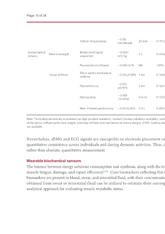
               Note: Technological maturity is evaluated as high (product available), medium (human validation available), and low (prototype only). Comfort is classified is evaluated as high, medium, and low based on the skin conformability
               of the device (influenced by size, weight, substrate stiffness and mechanical structure design). sEMG: Surface electromyography; Mb: myoglobin; CK: creatine kinase; IL-6: Interleukin-6; TGF-β: transforming growth factor-β; NA:
               not available.

               Nevertheless, sEMG and ECG signals are susceptible to electrode placement variability, cross-talk, and motion artifacts, which may affect reproducibility and
               quantitative consistency across individuals and during dynamic activities. Thus, current bioelectrical wearables are better suited for relative, qualitative monitoring
               rather than absolute, quantitative measurement.

               Wearable biochemical sensors
               The balance between energy substrate consumption and synthesis, along with the level of inflammatory response, constitutes a key dimension for assessing the extent of
               muscle fatigue, damage, and repair efficiency [105] . Core biomarkers reflecting this balance encompass reserved glycogen, lactate, LDH, UN, Mb, CK and IL-6. These
               biomarkers are present in blood, sweat, and interstitial fluid, with their concentration dynamics exhibiting significant cross-correlations [106,107] . Therefore, measurements
               obtained from sweat or interstitial fluid can be utilized to estimate their corresponding levels in blood, thereby providing a non‑invasive or minimally invasive
               analytical approach for evaluating muscle metabolic status.

