


Page 136 - Read Online
P. 136
Page 14 of 19                                                        Chen et al. Soft Sci. 2026, 6, 48


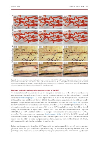
               Figure 5. Magnetic navigation and angioplasty demonstration of the MBF. (A) The MBF successfully navigates complex and tortuous
               cerebral arteries such as the Circle of Willis (CW), right internal carotid artery (ICA), and right middle cerebral artery (MCA) in a 3D
               cerebrovascular phantom; (B) Magnetic navigation of the MBF in an ex vivo porcine placenta; (C) Phase-change expansion of the MBF via
               microwave heating. MBF: Magnetic balloon fiberbot; 3D: three-dimensional.


               Magnetic navigation and angioplasty demonstration of the MBF
               To comprehensively evaluate the magnetic navigation performance of the MBF, we conducted a
               demonstration using a 3D-printed cerebrovascular phantom that replicates the intricate human cerebral
               arteries [Supplementary Figure 13], including the Circle of Willis (CW), the right internal carotid artery
               (ICA), and the right middle cerebral artery (MCA). Guided by external magnetic fields, the MBF successfully
               navigated through complex and tortuous branches. The navigation sequence, shown in Figure 5A, highlights
               the MBF’s ability to access small and narrow arterial branches. At 00:09, the MBF passed the curved ICA
               with a diameter of 3 mm. At 00:22, it successfully entered CW. Remarkably, at 00:38, the MBF navigated
               through an extremely narrow segment with a diameter of 1.6 mm. After that, MBF reached the M2 segment
               of the MCA at 00:47. The MBF continued to navigate seamlessly into even smaller arteries of MCA
               thereafter. The time-sequenced images and Supplementary Video 3 highlight the MBF’s smooth and
               consistent movement, even in highly curved and confined segments of the phantom. This demonstration
               underscores the MBF’s excellent navigation capabilities in small and tortuous blood vessels effectively,
               offering a promising solution for angioplasty in small arteries.


               Following the demonstration of the MBF’s magnetic navigation capabilities in a 3D cerebrovascular
               phantom, we further performed biocompatibility testing and an ex vivo angioplasty demonstration in a
               porcine placenta model to assess its feasibility in a biologically relevant vascular environment. Cytotoxicity

