
Page 143 - Read Online
P. 143
Page 6 of 15 Li et al. Soft Sci 2023;3:22 https://dx.doi.org/10.20517/ss.2023.11
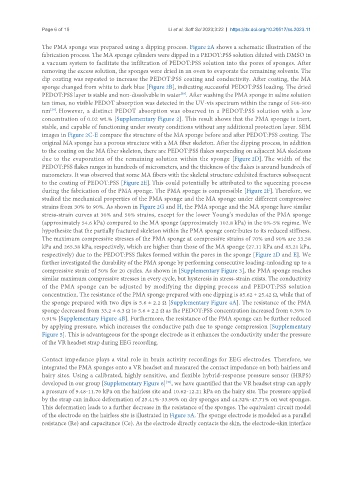
The PMA sponge was prepared using a dipping process. Figure 2A shows a schematic illustration of the
fabrication process. The MA sponge cylinders were dipped in a PEDOT:PSS solution diluted with DMSO in
a vacuum system to facilitate the infiltration of PEDOT:PSS solution into the pores of sponges. After
removing the excess solution, the sponges were dried in an oven to evaporate the remaining solvents. The
dip coating was repeated to increase the PEDOT:PSS coating and conductivity. After coating, the MA
sponge changed from white to dark blue [Figure 2B], indicating successful PEDOT:PSS loading. The dried
PEDOT:PSS layer is stable and non-dissolvable in water . After washing the PMA sponge in saline solution
[38]
ten times, no visible PEDOT absorption was detected in the UV-vis spectrum within the range of 500-800
nm . However, a distinct PEDOT absorption was observed in a PEDOT:PSS solution with a low
[39]
concentration of 0.02 wt.% [Supplementary Figure 2]. This result shows that the PMA sponge is inert,
stable, and capable of functioning under sweaty conditions without any additional protection layer. SEM
images in Figure 2C-E compare the structure of the MA sponge before and after PEDOT:PSS coating. The
original MA sponge has a porous structure with a MA fiber skeleton. After the dipping process, in addition
to the coating on the MA fiber skeleton, there are PEDOT:PSS flakes suspending on adjacent MA skeletons
due to the evaporation of the remaining solution within the sponge [Figure 2D]. The width of the
PEDOT:PSS flakes ranges in hundreds of micrometers, and the thickness of the flakes is around hundreds of
nanometers. It was observed that some MA fibers with the skeletal structure exhibited fractures subsequent
to the coating of PEDOT:PSS [Figure 2E]. This could potentially be attributed to the squeezing process
during the fabrication of the PMA sponge. The PMA sponge is compressible [Figure 2F]. Therefore, we
studied the mechanical properties of the PMA sponge and the MA sponge under different compressive
strains from 30% to 90%. As shown in Figure 2G and H, the PMA sponge and the MA sponge have similar
stress-strain curves at 30% and 50% strains, except for the lower Young’s modulus of the PMA sponge
(approximately 54.6 kPa) compared to the MA sponge (approximately 102.8 kPa) in the 0%-5% regime. We
hypothesize that the partially fractured skeleton within the PMA sponge contributes to its reduced stiffness.
The maximum compressive stresses of the PMA sponge at compressive strains of 70% and 90% are 33.58
kPa and 265.38 kPa, respectively, which are higher than those of the MA sponge (27.11 kPa and 83.21 kPa,
respectively) due to the PEDOT:PSS flakes formed within the pores in the sponge [Figure 2D and E]. We
further investigated the durability of the PMA sponge by performing consecutive loading-unloading up to a
compressive strain of 50% for 20 cycles. As shown in [Supplementary Figure 3], the PMA sponge reaches
similar maximum compressive stresses in every cycle, but hysteresis in stress-strain exists. The conductivity
of the PMA sponge can be adjusted by modifying the dipping process and PEDOT:PSS solution
concentration. The resistance of the PMA sponge prepared with one dipping is 85.62 ± 25.42 Ω, while that of
the sponge prepared with two dips is 5.6 ± 2.2 Ω [Supplementary Figure 4A]. The resistance of the PMA
sponge decreased from 33.2 ± 6.3 Ω to 5.6 ± 2.2 Ω as the PEDOT:PSS concentration increased from 0.39% to
0.91% [Supplementary Figure 4B]. Furthermore, the resistance of the PMA sponge can be further reduced
by applying pressure, which increases the conductive path due to sponge compression [Supplementary
Figure 5]. This is advantageous for the sponge electrode as it enhances the conductivity under the pressure
of the VR headset strap during EEG recording.
Contact impedance plays a vital role in brain activity recordings for EEG electrodes. Therefore, we
integrated the PMA sponges onto a VR headset and measured the contact impedance on both hairless and
hairy sites. Using a calibrated, highly sensitive, and flexible hybrid-response pressure sensor (HRPS)
[40]
developed in our group [Supplementary Figure 6] , we have quantified that the VR headset strap can apply
a pressure of 9.48-11.70 kPa on the hairless site and 10.62-12.21 kPa on the hairy site. The pressure applied
by the strap can induce deformation of 25.41%-33.90% on dry sponges and 44.32%-47.71% on wet sponges.
This deformation leads to a further decrease in the resistance of the sponges. The equivalent circuit model
of the electrode on the hairless site is illustrated in Figure 3A. The sponge electrode is modeled as a parallel
resistance (Re) and capacitance (Ce). As the electrode directly contacts the skin, the electrode-skin interface