
Page 65 - Read Online
P. 65
Page 6 of 29 Aguiar. Rare Dis Orphan Drugs J 2024;3:13 https://dx.doi.org/10.20517/rdodj.2023.56
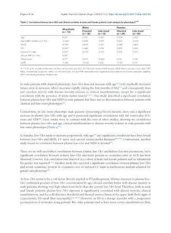
Table 2. Correlation between lyso-Gb3 and clinical variables in male and female patients (sub-analysis by phenotype) [46]
Males Females
All patients
(n = 73) Classical Late-onset Classical Late-onset
(n = 18) (n = 11) (n = 29) (n = 15)
Age 0.001 0.325 -0.247 0.309 0.082
Age at ERT initiation (n = 50) -0.466* 0.088 -0.071 0.230 -0.100
MSSI 0.538* 0.458 -0.251 0.441* 0.404
FIPI 0.383* 0.424 -0.196 0.259 0.363
Indexed LV mass 0.353* 0.776* -0.327 0.303 0.045
NT-pro BNP (n = 24) 0.247 - - - -
Albuminuria 0.317* 0.079 -0.600 0.133 0.380
eGFR -0.168 -0.066 0.345 -0.125 0.015
*P < 0.05; ρ for correlation between variables and plasma lyso-Gb3. ERT: Enzyme replacement therapy; MSSI: Mainz severity score index; FIPI:
Fabry international prognostic index; LV: left ventricular; NT-pro BNP: amino-terminal fragment of the prohormone of brain natriuretic peptide;
eGFR: estimated glomerular filtration rate.
In male patients with classical phenotype, lyso-Gb3 does not increase with age (with markedly increased
[49]
values even in neonates, which increases rapidly during the first months of life) and consequently does
[58]
not correlate strictly with disease severity indexes or clinical manifestations, except for a significant
correlation with the presence of white matter lesions [1,40,41] . One study described a significant correlation
between plasma lyso-Gb3 and MSSI in male patients, but there was no discrimination between patients with
classical and late-onset phenotypes .
[43]
Contrariwise, in late-onset phenotype male patients (presenting p.N215S variant), there was a significant
increase in plasma lyso-Gb3 with age and it presented significant correlations with left ventricular (LV)
mass and GFR ; these results were in contrast with the ones of other studies, showing no correlations
[48]
between plasma lyso-Gb3 and age, clinical manifestations or disease severity indexes in male patients with
[46]
late-onset phenotypes [Table 2] .
In females, lyso-Gb3 tends to increase progressively with age and significant correlations have been found
[49]
between lyso-Gb3 and MSSI, LV mass, and carotid intima-media thickness [40,41,59] . Contrariwise, another
study found no correlation between plasma lyso-Gb3 and MSSI in females .
[43]
There are no well-established correlations between plasma lyso-Gb3 and kidney function parameters, but a
significant correlation between urinary lyso-Gb3 and urine protein-to-creatinine ratio or ACR has been
observed; however, this correlation was observed in a cohort of male and female patients and no adjustment
for gender was reported [1,33] . Another study also reported a significant correlation between plasma lyso-Gb3
and serum creatinine, protein-to-creatinine ratio or indexed LV mass in multivariate analysis adjusted for
[49]
gender and phenotype .
As lyso-Gb3 seems to be a risk factor directly implied in FD pathogenesis, lifetime exposure to plasma lyso-
Gb3 (estimated product of lyso-Gb3 concentration by age) should correlate better with disease (mainly in
male patients, showing very high values from birth) than the current lyso-Gb3 level. Therefore, both in male
and female patients, plasma lyso-Gb3 exposure is significantly correlated with disease severity, clinical
manifestations, and the cold detection threshold and thermal sensory limen of the upper limb (both signs of
characteristic FD small fiber neuropathy) [1,41,48,60] . However, as FD is a storage disorder with a progressive
accumulation of substrates along patients’ life, older patients tend to have more severe manifestations; thus,