
Page 612 - Read Online
P. 612
Page 4 of 13 Ziai et al. Plast Aesthet Res 2020;7:53 I http://dx.doi.org/10.20517/2347-9264.2020.151
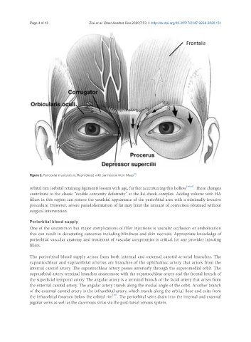
Figure 2. Periocular musculature. Reproduced with permission from Maas [11]
orbital rim (orbital retaining ligament) loosen with age, further accentuating this hollow [15,16] . These changes
contribute to the classic “double convexity deformity” at the lid-cheek complex. Adding volume with HA
fillers in this region can restore the youthful appearance of the periorbital area with a minimally invasive
procedure. However, severe pseudoherniation of fat may limit the amount of correction obtained without
surgical intervention.
Periorbital blood supply
One of the uncommon but major complications of filler injections is vascular occlusion or embolization
that can result in devastating outcomes including blindness and skin necrosis. Appropriate knowledge of
periorbital vascular anatomy and treatment of vascular compromise is critical for any provider injecting
fillers.
The periorbital blood supply arises from both internal and external carotid arterial branches. The
supratrochlear and supraorbital arteries are branches of the ophthalmic artery that arises from the
internal carotid artery. The supratrochlear artery passes anteriorly through the superomedial orbit. The
supraorbital artery terminal branches anastomose with the supratrochlear artery and the frontal branch of
the superficial temporal artery. The angular artery is a terminal branch of the facial artery that arises from
the external carotid artery. The angular artery travels along the medial angle of the orbit. Another branch
of the external carotid artery is the infraorbital artery, which travels along the orbital floor and exits from
[17]
the infraorbital foramen below the orbital rim . The periorbital veins drain into the internal and external
jugular veins as well as the cavernous sinus via the post-tarsal venous system.