
Page 617 - Read Online
P. 617
Ziai et al. Plast Aesthet Res 2020;7:53 I http://dx.doi.org/10.20517/2347-9264.2020.151 Page 9 of 13
Figure 5. Hyaluronic acid injection to the superior sulcus. The needle or cannula should enter along the superior orbital rim at a 30°
angle. After the needle has reached the bone, it should be slightly withdrawn to the preperiosteal space to inject into the suborbicularis
plane. Adapted with permission from Looi et al. [31]
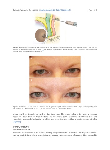
A
B
Figure 6. Combination of hyaluronic acid injection into the glabellar rhytids with chemodenervation: (A) pre-injection; and (B) two
months after the glabellar injection of 0.1 mL on the right and 0.2 mL on the left of Restylane-L
with a low G’ are typically required to efface these lines. The senior author prefers using a 30-gauge
needle with bevel down for these injections. The filler should be injected in the subcutaneous plane and
immediately massaged after injection to achieve an even contour and avoid early-onset nodules or visibility
[Figure 6].
COMPLICATIONS
Vascular occlusion
Vascular occlusion is one of the most devastating complications of filler injections. In the periocular area,
this can result in intra-arterial embolization or vascular compression and subsequent vision loss or skin