
Page 611 - Read Online
P. 611
Ziai et al. Plast Aesthet Res 2020;7:53 I http://dx.doi.org/10.20517/2347-9264.2020.151 Page 3 of 13
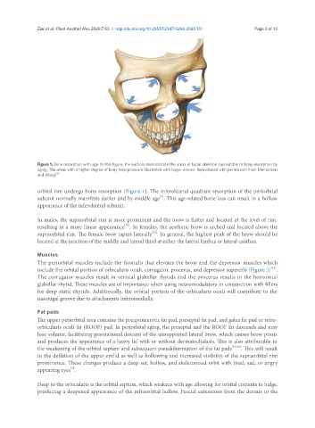
Figure 1. Bone resorption with age. In this figure, the authors demonstrate the areas of facial skeleton susceptible to bony resorption by
aging. The areas with a higher degree of bony resorption are illustrated with larger arrows. Reproduced with permission from Mendelson
and Wong [3]
orbital rim undergo bony resorption [Figure 1]. The inferolateral quadrant resorption of the periorbital
[3]
subunit normally manifests earlier and by middle age . This age-related bone loss can result in a hollow
appearance of the inferolateral subunit.
In males, the supraorbital rim is more prominent and the brow is flatter and located at the level of rim,
[10]
resulting in a more linear appearance . In females, the aesthetic brow is arched and located above the
[11]
supraorbital rim. The female brow tapers laterally . In general, the highest peak of the brow should be
located at the junction of the middle and lateral third at either the lateral limbus or lateral canthus.
Muscles
The periorbital muscles include the frontalis that elevates the brow and the depressor muscles which
[12]
include the orbital portion of orbicularis oculi, corrugator, procerus, and depressor supercilii [Figure 2] .
The corrugator muscles result in vertical glabellar rhytids and the procerus results in the horizontal
glabellar rhytid. These muscles are of importance when using neuromodulators in conjunction with fillers
for deep static rhytids. Additionally, the orbital portion of the orbicularis oculi will contribute to the
nasojugal groove due to attachments inferomedially.
Fat pads
The upper periorbital area contains the preaponeurotic fat pad, preseptal fat pad, and galea fat pad or retro-
orbicularis oculi fat (ROOF) pad. In periorbital aging, the preseptal and the ROOF fat descends and may
lose volume, facilitating gravitational descent of the unsupported lateral brow, which causes brow ptosis
and produces the appearance of a heavy lid with or without dermatochalasis. This is also attributable to
the weakening of the orbital septum and subsequent pseudoherniation of the fat pads [13,14] . This will result
in the deflation of the upper eyelid as well as hollowing and increased visibility of the supraorbital rim
prominence. These changes produce a deep-set, hollow, and skeletonized orbit with tired, sad, or angry
[1]
appearing eyes .
Deep to the orbicularis is the orbital septum, which weakens with age allowing for orbital contents to bulge,
producing a deepened appearance of the infraorbital hollow. Fascial extensions from the dermis to the