
Page 510 - Read Online
P. 510
Mirastschijski et al. Plast Aesthet Res 2020;7:45 I http://dx.doi.org/10.20517/2347-9264.2020.44 Page 9 of 13
A B
C D
E F
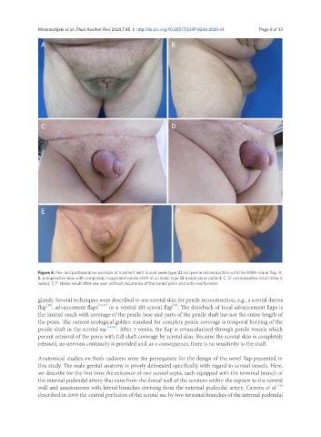
Figure 4. Pre- and postoperative example of a patient with buried penis type III and penile reconstruction with the MiRA island flap. A,
B: preoperative view with completely invaginated penile shaft of a classic type III buried penis patient; C, D: postoperative result after 4
weeks; E, F: stable result after one year without recurrence of the buried penis and with neo-foreskin
glands. Several techniques were described to use scrotal skin for penile reconstruction, e.g., a scrotal dartos
[22]
[20]
flap , advancement flaps [13,21] or a ventral slit scrotal flap . The drawback of local advancement flaps is
the limited reach with coverage of the penile base and parts of the penile shaft but not the entire length of
the penis. The current urological golden standard for complete penile coverage is temporal burying of the
penile shaft in the scrotal sac [14,23] . After 3 weeks, the flap is revascularized through penile vessels which
permit retrieval of the penis with full shaft coverage by scrotal skin. Because the scrotal skin is completely
released, no nervous continuity is provided and, as a consequence, there is no sensitivity to the shaft.
Anatomical studies on fresh cadavers were the prerequisite for the design of the novel flap presented in
this study. The male genital anatomy is poorly delineated specifically with regard to scrotal vessels. Here,
we describe for the first time the existence of two scrotal septa, each equipped with the terminal branch of
the internal pudendal artery that runs from the dorsal wall of the scrotum within the septum to the ventral
[16]
wall and anastomoses with lateral branches deriving from the external pudendal artery. Carrera et al.
described in 2009 the central perfusion of the scrotal sac by two terminal branches of the internal pudendal