
Page 506 - Read Online
P. 506
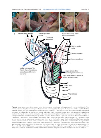
Mirastschijski et al. Plast Aesthet Res 2020;7:45  I  http://dx.doi.org/10.20517/2347-9264.2020.44                           Page 5 of 13

                A                     B                                 C                 D


                 E


               Figure 2. Septal anatomy with demonstration of the dual presence of scrotal septa including an arch forming terminal branch of the
               internal pudendal artery that is running from the dorsal to the ventral aspect of the septum. A: lateral view with the flap folded behind
               the septa. Arrows depicting the septal artery of each septum; B: close-up of A with the septal arteries (thick arrows) and anastomoses
               with lateral scrotal branches derived from the external pudendal artery (thin arrows); C: septal artery with anastomoses to the lateral
               border of the flap; D: close-up of the left septum with upper arrow depicting the septal artery. Lower arrow pointing to the artery of
               the right septum that is visible shining through the tissue layer of the left septum; E: graphical overview of the vascular anatomy of
               the scrotum. The scrotum is vascularized by the anterior, lateral and posterior branches of different arteries. The main vessels of the
               scrotum are the internal and external pudendal artery. After leaving the Alcock channel (ischiorectal fascia, blue long arrow), the
               internal pudendal vessels (1) divide into the rectal (not shown) and perineal artery (2). The dorsal scrotal branches (3) originate from
               the perineal artery which continues in the midline as the septal artery (4, red arrow). Further branches of each internal pudendal artery
               are the central arteries - providing the ipsilateral cavernosal bodies (5) and the corpus spongiosum (6) - and the dorsal penile artery (7).
               The anterior (8) and lateral (9) scrotal branches derive from the external pudendal artery originating from the femoral artery. Of note,
               the pudendal nerve (10, green arrow) accompanies the internal pudendal artery and its branches throughout the scrotum and penis