
Page 508 - Read Online
P. 508
Mirastschijski et al. Plast Aesthet Res 2020;7:45 I http://dx.doi.org/10.20517/2347-9264.2020.44 Page 7 of 13
A B C D
E F
G H
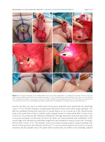
Figure 3. The surgical technique of the midline raphe scrotal artery flap is presented. A: preoperative markings; B: flap harvest and
positioning around the penile shaft with complete coverage; C-F: the terminal branch of the pudendal artery runs in an arch-formed
way from the dorsal plane of the septum to the ventral side. The black arrow depicts the artery (D-F), the white arrow depicts the
concomitant nerve (E); G, H: postoperative result after wound closure. Visualization of the septal artery by diaphanoscopy in F
incision, the flap was raised on both septal arteries preserving both septa underneath the island flap
[Figure 3D-F]. Vascular branches anastomosing with lateral vessels were sealed using LigaClips®. The
flap was mobilized, freeing both septa from its caudal fixation in the scrotum up to the cranial fixation
point at the penile base so that it could be easily wrapped around the entire penile shaft. Closure of the
scrotal sac was performed after bilateral orchidopexy, thorough hemostasis and drain placement. Due
to excessive and elastic scrotal tissue, the donor site defect was closed primarily after mobilization of the
wound edges with subcutaneous absorbable single knots and intradermal running sutures after inserting a
Penrose drain [Figure 3G-H]. The prepubic wound was closed according to plastic-surgical standards with
[18]
fascial anchoring sutures after Baroudi and Ferreira to avoid seroma formation and recurrence of penile
retraction into the prepubic tissue. For penile shaft reconstruction, the MiRA scroti island flap, pedicled