
Page 182 - Read Online
P. 182
Patel et al. Plast Aesthet Res 2020;7:18 I http://dx.doi.org/10.20517/2347-9264.2019.15 Page 3 of 7
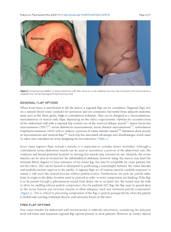
Figure 1. Fasciocutaneousdefect of lateral abdominal wall after resection of an irradiated sarcoma. Reconstructed with fasciocutaneous
propeller flap from lumbar segmental perforating vessel
REGIONAL FLAP OPTIONS
When local tissue is insufficient to fill the defect, a regional flap can be considered. Regional flaps rely
on a named blood vessel (pedicle) for perfusion and are commonly harvested from adjacent anatomic
areas such as the chest, groin, thigh or contralateral abdomen. They can be designed as a fasciocutaneous,
myocutaneous or muscle-only flaps, depending on the defect requirements. Options for reconstruction
[7]
of the abdominal wall with a regional flap include use of the external oblique muscle , tensor fascia lata
[10]
[8,9]
myocutaneous (TFL) , rectus abdominis myocutaneous, rectus femoris myocutaneous , anterolateral
[11]
thighfasciocutaneous (ALT) with or without a portion of vastus lateralis muscle , latissimus dorsi muscle
[12]
or myocutaneous and omental flaps . Each flap has associated advantages and disadvantages which must
be taken into consideration when designing the reconstruction [Table 1].
Since many regional flaps include a muscle, it is important to consider donor morbidity. Although a
contralateral rectus abdominis muscle can be used to reconstruct a portion of the abdominal wall, the
weakness and hernia potential incurred by moving this muscle may preclude its use. Similarly, the rectus
femoris can be used to reconstruct the infraumbilical abdomen, however using this muscle may limit the
terminal fifteen degrees of knee extension of the donor leg; this may be acceptable for some patients but
not for others. This can be limited or eliminated by performing a tenorrhaphy between the vastus lateralis
and medialis tendons superior to the patella. A regional flap’s arc of rotation must be carefully measured to
ensure it will reach the desired location without pedicle tension. Furthermore, the path the pedicle takes
from its origin to the defect location must be planned in order to avoid compression and kinking. If the flap
is to be passed through a subcutaneous tunnel from donor site to recipient site, the tunnel must be wide
to allow for swelling without pedicle compression. For the pedicled ALT flap, the flap must be passed deep
to the rectus femoris and sartorius muscles to allow adequate reach and minimize pedicle compression
[Figure 2]. This is critical in preventing compression of the flap or pedicle postoperatively when the patient
is mobile and exerting rotational, flexion, and extension forces on the torso.
FREE FLAP OPTIONS
Free tissue transfer for abdominal wall reconstruction is relatively uncommon, considering the adequate
local soft tissue and numerous regional flap options present in most patients. However, in certain clinical