
Page 184 - Read Online
P. 184
Patel et al. Plast Aesthet Res 2020;7:18 I http://dx.doi.org/10.20517/2347-9264.2019.15 Page 5 of 7
A B C
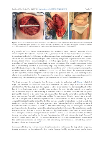
Figure 3. 20 cm × 20 cm full thickness defect of abdominal wall with exposed viscera (A). Myofascial defect reconstructed with a bridged
bioprosthetic mesh and free anterolateral thigh myocutaneous flap anastomosed to the right deep inferior epigastric vessels (B). Healed
and viable reconstruction 1 month postoperatively; anterolateral thigh flap donor site closed with skin graft (C)
2
flap provides well-vascularized soft tissue to resurface a defect of up to a 1500 cm . However, it bears
mentioning that if the latissimus muscle is included, donor site morbidity must be considered as it relates to
the weakened abdominal wall. Patients who have decreased core muscle strength as a result of a composite
abdominal wall resection will depend upon upper extremity strength and range of motion more so than
is usual. Simple actions - such as rising from a seated or supine position - extensively utilize the trunk
musculature. If core strength has been reduced, the upper extremities will be needed to compensate for the
lack of trunk stability. Therefore, in patients requiring a large free flap, preference should be given to lower-
morbidity fasciocutaneous flap options, such as the thoracodorsal artery perforator, scapular/parascapular,
or fasciocutaneous subtotal thigh. In addition, if a posterior trunk flap is chosen, the patient must undergo
an intra-operative position change to elevate the flap on the posterior chest wall, then another position
change to supine to inset the flap. The surgeon must be aware of these logistical issues, since intraoperative
repositioning adds complexity, prolongs the operative duration, and extends flap ischemia time.
The thigh represents the mainstay for free flap donor sites for the abdominal wall [Figure 3]. Pedicled
thigh flaps can reach the infraumbilical abdomen, however if the defect is larger or located outside the
arc of rotation, the thigh flap must be designed as a free tissue transfer. The descending branch of the
lateral circumflex femoral system provides blood supply to the vastus lateralis, rectus femoris muscles
and anterolateral thigh skin. The ascending or transverse branch of the lateral circumflex femoral system
provides blood supply to the tensor fascia lata muscle. These flaps can be harvested as muscle only flaps
or as myocutaneous flaps with overlying skin paddles. The anterolateral thigh flap is particularly versatile
and can be designed with or without a segment of vastus lateralis muscle. The tensor fascia lata flap can be
designed to include the distal fascia of the iliotibial tract and a smaller proximal skin paddle if needed; this
fascia can be used to reconstruct the fascial component of an abdominal wall defect, providing vascularized
tissue for reconstruction in lieu of mesh [9,16] . The anteromedial thigh flap can be designed on medial
perforators from the descending branch of the lateral circumflex femoral system. The rectus femoris muscle
is more commonly designed as a muscle flap however a skin island can be included over the central muscle
[13]
when appropriate sized cutaneous perforators are present . Similar to the subscapular system, the lateral
femoral circumflex vessels allow for chimeric flap design, i.e., ALT with anteromedial thigh flaps, ALT
with TFL, vastus lateralis with TFL. For massive abdominal wall defects the vastus lateralis, tensor fascia
lata, and the rectus femoris can be harvested with all overlying skin territory as a subtotal thigh flap for
[13]
increased volume and skin coverage .
Easily accessible free flap recipient vessels along the abdominal wall include the deep inferior epigastric,
internal mammary, and deep circumflex iliac vessels. However, healthy recipient vessels may be absent