
Page 117 - Read Online
P. 117
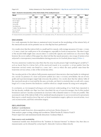
Page 4 of 5                             Muñoz-Jimenez et al. Plast Aesthet Res 2018;5:14  I  http://dx.doi.org/10.20517/2347-9264.2018.04

               Table 1. Anatomic characteristics of the inferior belly of the omohyoid muscle
                Model                  # 1          # 2           # 3          # 4          # 5      Mean
                                    Left  Right  Left  Right  Left  Right   Left  Right  Left  Right
                Vascular pedicle length  15  17  28     -      13    21     23    -      20     42    22.3
                Nerve length        46     17    37     -      26    21     31    -      25     20    27.8
                Flap length         67     90    85     -      110   100    84    -      100    110   93.2
                Flap width          20     16    16     -     6      11     11    -      12     11    12.8
                Flap thickness      8      7     6      -     6      7      7     -      9      12    7.7
                Tendon-Pedicle distance  19  19  27     -   No central tendon  25  -     75     53    27.2
                Source vessel       SC     SC    SC     -     SC     SC     SC    -      TC     TC
                Main nerve          AC     AC    AC     -     AC     AC     AC    -      AC     AC
               All measurements are expressed in millimeters (mm). SC: subclavian artery; TC: transverse cervical artery; AC: ansa cervicalis

               DISCUSSION
               Our work represents the first time an anatomical study focused on the morphology of the inferior belly of
               the omohyoid muscle and its potential use as a free flap has been performed.

               Our results show that the inferior belly is a small and thin muscle, with average measures of 93 mm × 12 mm
               × 7.5 mm, a trait that could prove to be advantageous, especially in facial reconstruction. The short length
               of the muscle’s belly might raise concern about its contractile capacity and usefulness as a functional flap;
               however the senior author of this paper has observed that the muscle presents a nice range of motion when
               subjected to transoperatory neurostimulation during procedures for brachial plexus injury [Video 1].

               Previous anatomical studies have described that the strap muscles present high morphological variability ,
                                                                                                        [2]
               and we found that the inferior belly of the omohyoid muscle is no exception: a minor pedicle from the
               transverse cervical vessels was found in 2 cases, and 1 case in which the main pedicle came from the
               transverse cervical artery, in lieu of the subclavian artery.


               The vascular pedicle of the inferior belly presents anatomical characteristics that may hinder its widespread
               use, mainly the presence of a short and narrow pedicle (22.3 mm × 0.8 mm), nevertheless, the use of vein
               grafts and supermicrosurgery might help overcome the problem. Our results show that the inferior belly of
               the omohyoid muscle is innervated by the ansa cervicalis, and that it is relatively short as well, with a mean
               length of 27.8 mm, however this limitation could be solved in a simple manner by using nerve autografts.


               In conclusion, as microsurgical techniques and anatomical understanding of our body have expanded in
               the last decade, multiple new flaps have been described; the use of supermicrosurgery has further pushed
               boundaries and now vascular anastomosis in structures with a diameter minor to 0.8 mm are possible. This
               anatomical study of the inferior belly of the omohyoid muscle proposes its use in reconstructive procedures
               that require a small myofunctional flap such as facial reanimation surgery, sphincter reconstruction, vocal
               cord reconstruction, and blink restoration surgery.


               DECLARATIONS
               Authors’ contributions
               Anatomical model dissection, data acquisition and analysis: Muñoz-Jimenez G
               Data analysis, paper writing, editing and translation: Telich-Tarriba JE, Palafox-Vidal D
               Project supervisor and anatomical model dissection: Cardenas-Mejia A

               Data source and availability
               Anatomical models were obtained from the “Institute de Ciencias Forenses” in Mexico City. Raw data on
               measurements can be obtained by contacting Dr. Alexander Cardenas-Mejia directly.