
Page 84 - Read Online
P. 84
Kumar et al. Reverse latissimus dorsi muscle flap for back
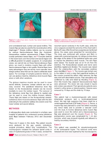
Figure 1: Latissimus dorsi muscle flap raised based on the Figure 2: Latissimus dorsi muscle inset given, donor site closed.
secondary pedicle Viability confirmed before closing the wound
and contralateral back, lumbar and sacral midline. This recurrent sacral cordoma in the fourth case, while the
muscle flap can also be used for the reconstruction of the onco surgeons excised the sarcoma of the chest wall in
diaphragm. Simple midline defects can be covered the fourth case. After the primary surgeries as mentioned
[3]
by various fasciocutaneous flaps like rhomboid, above, the cases were presented for reconstruction.
transposition, rotation, advancement, Z flaps, S flaps, The dura was reinforced with sutures and fibrin in
lumbar perforator based flaps, etc. Complex defects cases with myelomeningocele. An oblique incision from
[2]
in the lower back over the lumbar and sacral region are the edge of the defect to the ipsilateral axilla was used
a difficult problem for plastic surgeons. In complicated to expose the latissimus dorsi muscle. The skin flaps
cases, we cannot rely on these fasciocutaneous flaps were raised. The muscle was cut at 10 cm from the
alone, we need to combine muscle flaps with either insertion. The thoracodorsal vessels and nerve were
fasciocutaneous flaps or skin grafts. Muscle flaps have identified, ligated and divided. The muscle was raised
many advantages: they are sturdy, more vascular, have from the undersurface until the secondary pedicles
alarge surface area that can be used to obliterate dead were identified [Figure 1]. The muscle was turned over
space. For coverage of complex posterior defects, we to the defect in such a way that superficial surface of
can use gluteus maxims, latissimus dorsi muscle flaps the muscle covered the defect. After turnover, the flap
alone or with other variations. was inset with absorbable sutures. Flap vascularity was
confirmed by color and distal tip bleeding [Figure 2].
The gluteus maximus muscle can be used to cover Over this muscle flap, adjacent fasciocutaneous flaps
sacral defects. However, lumbar defects are not as were advanced. The donor site was closed in layers
easy covered. The ipsilateral latissimus dorsi flap and a drain was placed. Post operatively patients were
based on the thoracodorsal vessels can be moved nursed in either prone or lateral position. Sutures were
medially to cover the lumbar region. The turnover of removed by 15 days and the drain by 10 days.
the latissimus dorsi flap from lateral to medial side
can be used to cover the midline thoracic defects. Case 1
These flaps are based on the thoracodorsal vessels. This patient had a myelomeningocele with raised
Latissimus dorsi musculocutaneous flaps with the skin intracranial pressure and a ventriculoperitoneal
extending to the posterior axillary line would cover the shunt. We had high suspicion that there might be a
lower lumbar and sacral defects. cerebrospinal fluid leak (CSF leak) [Figure 3]. Hence, a
muscle flap was planned to cover the defect including
METHODS any minor leaks and to provide a vascular cover
over the dura. A reverse latissimus dorsi muscle flap
A retrospective study was conducted in a tertiary care was used to cover the defect and a fasciocutaneous
hospital of patients who underwent reverse latissimus flap was advanced over the muscle [Figure 2]. The
dorsi flaps between February 2012 and December postoperative course was complicated by 1 cm skin
2016. necrosis, which was dressed regularly and allowed to
heal by secondary intention.
There are 5 cases in this study. The patient records
were analyzed for the age, gender, indications, Case 3
surgical procedure, defect size, complications. The This patient was operated for amyelomeningocele
neurosurgeons released the tethered spinal cords in of the lumbosacral region. The tethered cords were
case of myelomeningocele in first 3 cases, excised the released and dura was repaired by the neurosurgeons.
Plastic and Aesthetic Research ¦ Volume 4 ¦ May 26, 2017 77