
Page 72 - Read Online
P. 72
Maher et al. Hypospadias: an algorithm for repair
METHODS 3 times a day. Both medications are continued until
the catheter is removed at 1 week post-operatively.
One hundred and two patients were operated upon Analgesics are given as and when required. We aim to
between 2009 and 2013; the senior author (M. Dalal) discharge patients on the second post-operative day.
performed all patients. Inclusion criteria included They are reviewed at 1 and 2 weeks postoperatively.
primary hypospadias in the paediatric population They then are reviewed at 3 months, and from then on
irrespective of age at the time of initial presentation yearly till school age.
in clinic. Exclusion criteria, was 1 adult with delayed
presentation of primary hypospadias, and 5 cases of One stage repair (without urethral plate
phimosis, thought to have concealed hypospadias, incision)
treated with circumcision only. The following steps are A subcoronal marking is made on the dorsal penile
common to both one stage and two stage procedures. surface and is continued ventrally to the edge of the
All patients were subjected to a general anaesthetic urethral plate. The ventral markings are made all
augmented with a caudal block; for prolonged post- around the edges of the urethral plate and around the
operative pain relief. After induction, co-amoxiclav anomalous urethral opening in a “U” shaped design.
antibiotic (Augmentin-GlaxoSmithKline) at a dose The ventral incision is made around the urethral plate,
of 30 mg/kg is given intravenously over 3-4 min. and care is taken when dissecting the ventral skin
Medical photography after obtaining the consent off the anomalous urethral opening, as the corpus
from the parents is a routine part of our practice. The spongiosum is deficient, and there is a chance of injury
photographs are taken in 2 views once the patient is to the urethra. The use of a urethral dilator to guide this
covered in surgical drapes. part of the dissection can be very helpful [Figure 2].
Ventral chordee encountered at this stage is corrected
The foreskin is retracted and all the smegma removed by degloving the penis. Glans flaps are dissected off
with a swab soaked in aqueous chlorhexidine, the corpora cavernosa with a number fifteen Beaver
after which the surgeon exchanges the gloves to blade (Beaver Visitec), to achieve a tension free ventral
commence surgery. A urethral dilator size 6/8-8/10 repair over the reconstructed neo-urethra. The dorsum
is passed after lubrication to ensure that there is an of the penis is degloved in the sub-Dartos plane, and
adequate urethral calibre. A polypropylene 5-0 suture
(Prolene-Ethicon) is passed on the dorsal surface
of the glans for retraction. An 8 French (Fr) urethral
catheter is secured at the base of the penis with a
haemostat as a tourniquet to facilitate a bloodless
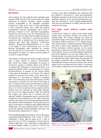
field. The microscope (Carl Zeiss Microscopy GmbH)
microscope is brought into the field at this stage to
aid in visualizing the anatomical landmarks as well as
carrying out the procedure under ×3.5-×6 magnification
[Figure 1]. A fine nib quill and ink are used to mark the
incision lines, the markings for which differ between
the one and two stage procedure. After the finishing
the surgery, the suture line is covered soft non-
adherent paraffin impregnated gauze (Jelonet-Smith
& Nephew) and sandwiched on the child’s abdomen
between two layers of low-adhesive perforated plastic
films (Melolin-Smith & Nephew). This is further secured
with a broad sheet of adhesive tape (Mefix-Mölnlycke
Healthcare). The same adhesive tape is then used to
secure the catheter and the paediatric urine bag. This
dressing is left undisturbed for 1 week.
Post-operatively patients are kept on oral co-amoxilcav
(Augmentin-GlaxoSmithKline) 0.25 mL/kg of 125/31
suspension 3 times daily for ages up to 1 year of
age and 5 mL of 125/31 suspension 3 times daily for
children older than 1 year. Oxybutynin is administered Figure 1: Use of microscope during surgery with the background
to prevent bladder spasm, at a dose of 1.25-2.5 mg, screen used for intraoperative teaching
Plastic and Aesthetic Research ¦ Volume 4 ¦ April 27, 2017 65