
Page 28 - Read Online
P. 28
Pérez et al. End-to-side neurorrhaphy for early reinnervation
A B C
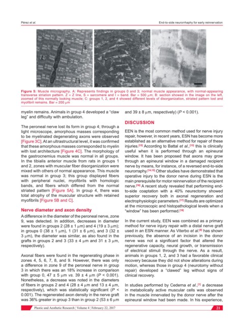
Figure 5: Muscle micrography. A: Represents findings in groups 0 and 3; normal muscle appearance, with normal-appearing
transverse striation pattern. Z = Z line, S = sarcomere and I = band. Bar = 500 mm; B: section showed in the image on the left,
zoomed of this normally looking muscle; C: groups 1, 2, and 4 showed different levels of disorganization, striated pattern lost and
myofibril remains. Bar = 200 mm
myelin remains. Animals in group 4 developed a “claw and 39 ± 8 mm, respectively) (P < 0.001).
leg” and difficulty with ambulation.
DISCUSSION
The peroneal nerve lost its form in group 4; through a
light microscope, amorphous masses corresponding EEN is the most common method used for nerve injury
to be myelinated degenerating axons were observed repair, however, in recent years, ESN has become more
[Figure 3C]. At an ultrastructural level, it was confirmed established as an alternative method for repair of these
that these amorphous masses corresponded to myelin injuries. [12] According to Battal et al., [13] this is clinically
with lost architecture [Figure 4C]. The morphology of useful when it is performed through an epineural
the gastrocnemius muscle was normal in all groups. window. It has been proposed that axons may grow
In the tibialis anterior muscle from rats in groups 1 through an epineural window in a damaged recipient
and 2, zones with muscular fiber disorganization were nerve by means, for instance, of a reversed end-to-side
mixed with others of normal appearance. This muscle neurorraphy. [14,15] Other studies have demonstrated that
was normal in group 3; this group displayed fibers operative injury to the donor nerve during ESN is the
with peripheral nuclei, myofibrils with homologic main prerequisite for motor reinnervation of the recipient
bands, and fibers which differed from the normal nerve. [16] A recent study revealed that performing end-
striated pattern [Figure 5A]. In group 4, there was to-side coaptation with a 40% neurectomy showed
total atrophy of the muscular structure with retained superior recovery both in axonal regeneration and
myofibrils [Figure 5B and C]. electrophysiologic parameters. [17] Results are optimized
at the microscopic and histopathological levels when a
Nerve diameter and axon density “window” has been performed. [18]
A difference in the diameter of the peroneal nerve, zone
9, was detected. In addition, decreases in diameter In the current study, EEN was combined as a primary
were found in groups 2 (28 ± 1 mm) and 4 (19 ± 3 mm); method for nerve injury repair with a distal nerve graft
[8]
in groups 0 (38 ± 1 mm), 1 (31 ± 6 mm), and 3 (32 ± used in an ESN manner. As Viterbo et al. has shown
3 mm), the diameter was similar, as also found in the previously, the absence of an incision in the donor
grafts in groups 2 and 3 (33 ± 4 mm and 31 ± 3 mm, nerve was not a significant factor that altered the
respectively). regenerative capacity, neural growth, or transmission
of electrical stimuli through the nerve. As a result,
Axonal fibers were found in the regenerating phase in animals in groups 1, 2, and 3 had a favorable clinical
zones 4, 5, 6, 7, 8, and 9. However, there was only recovery because they did not show alterations during
a difference in zone 9 of the peroneal nerve in group motion, whereas those in group 4 (neurotomy without
3 in which there was an 18% increase in comparison repair) developed a “clawed” leg without signs of
with group 0, 47 ± 5 mm vs. 39 ± 4 mm (P < 0.001). clinical recovery.
Nonetheless, a decrease was noted in the diameters
of fibers in groups 2 and 4 (28 ± 4 mm and 13 ± 4 mm, In studies performed by Cederna et al., a decrease
[1]
respectively), which was statistically significant (P < in metabolically active muscular cells was observed
0.001). The regenerated axon density in the nerve graft in the muscle innervated by the donor nerve after the
was 36% greater in group 3 than in group 2 (53 ± 6 mm epineural window had been made. In his experience,
Plastic and Aesthetic Research ¦ Volume 4 ¦ February 22, 2017 21