
Page 105 - Read Online
P. 105
Keren et al. NMR for human fat graft assessment
INTRODUCTION without the difficulties of manual collection of the
dispersed small fat droplets at the end of the study.
Autologous fat transplantation for soft-tissue
augmentation has become increasingly popular in METHODS
recent years. For more than a century fat grafting has
been used for facial contouring, breast augmentation, Isolation and preparation of human fat tissue
breast reconstruction after mastectomies, post- Fat was harvested from the thigh of a 50-year-old
traumatic deformities, congenital anomalies and woman undergoing suction-assisted lipectomy under
burn injuries. [1-3] general anesthesia. Prior to commencement of the
procedure, the areas of aspiration were injected with a
Fat tissue is abundant, readily available, inexpensive, local anesthesia solution containing lidocaine (0.5%)
host compatible, associated with low morbidity and and adrenaline in order to decrease bleeding during
can be harvested easily and repeatedly. However, fat aspiration and relieve pain after the procedure.
according to the literature, there is a varied overall The fat was aspirated using a 14-gauge 3-hole blunt
survival rate of the fat graft in the range of 20-70%. [4-6] cannula, and then processed under sterile conditions
for subsequent grafting into nude mice within 2 h of
Aspirated fat tissue used for autologous fat collection. Following aspiration, the fat-containing syringe
transplantation is devoid of blood microvessels which underwent 2 rounds of centrifugation (1,200 rpm, 10 min
have been destroyed during aspiration and removed at room temperature) and then was placed vertically
during processing prior to injection. Therefore, the fat for 10 min. [14-18]
tissue that is injected into a recipient is considered
to be an ischemic fat cell mass. During the early Between centrifugations, the three different layers
period following transplantation, the fat transplant (oil, adipose and fluid) were separated. After the
exists under hypoxic and hypo-nutritional conditions. last centrifugation, the adipose layer was collected
Revascularization fail to be initiated in this early and loaded into 2 mL syringes. All procedures were
period, apoptosis ensues and results in late fat performed under sterile conditions. [19]
cell degeneration, low viability and ultimately fat
resorption. [7] The participant gave her written informed consent, and
the study was reviewed and approved by the Helsinki
In order to maximize the surface area and hence committee of the Rambam Health Care Campus and
exposure to blood supply of the graft, surgeons now the institutional review board of the Technion Animal
inject very small aliquots of fat grafts into multiple Care and Use Committee.
subcutaneous tunnels (Coleman’s technique). [8]
Animals and study design
We previously developed a novel animal model in The study was composed of nine 7-week-old female
which human fat was grafted into the scalp area of CD-1 nude mice (Envigo, Jerusalem, Israel). Seven
nude mice. [9-13] This model allowed investigation of the mice received transplants of human fat, while two mice
mechanisms of fat absorbtion and exploration of the
efficacy of new compounds, with potential to increase served as controls without human fat transplantation.
the vasculature and viability of fat grafts. [14-18] In this The animals were housed in a specific pathogen-free
previous model, fat was grafted as a bolus to allow room, with 1 animal per cage in a room with an artificial
ease of collection and analysis.
12 h light/dark cycle at a constant temperature range
However, a bolus has a relatively small surface area (24 ± 2 ℃) and relative humidity (55 ± 10%). The mice
in contact with vascularized tissue, and therefore
the center of the graft suffers from higher rates of
ischemia, necrosis and resorption. Therefore, a new
and more clinically relevant animal model needed to
be developed.
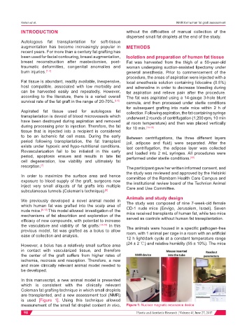
In this manuscript, a new animal model is presented
which is consistent with the clinically relevant
Coleman fat grafting technique in which small droplets
are transplanted, and a new assessment tool (NMR)
is used [Figure 1]. Using this technique allowed
measurement of the small fat droplet content in vivo, Figure 1: Nuclear magnetic resonance device
98 Plastic and Aesthetic Research ¦ Volume 4 ¦ June 27, 2017