
Page 61 - Read Online
P. 61
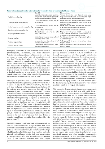
Table 3: Free tissue transfer alternatives for reconstruction of anterior skull base defects
                                         Benefits                    Disadvantages
                                         Versatile, reliable donor site anatomy,   Visible  donor site  scar.  Cannot  cover  very
          Radial forearm free flap       long pedicle length, size is appropriate   large skull base defects. Shorter hospital stays
                                         for most anterior skull base defects  compared to other free tissue transfers
                                         Consistent vascular pedicle and its   Larger donor site defect, greater risk of seroma,
          Latissimus dorsi muscle free flap  large muscle            may result in redundant bulky tissue in an anterior
                                                                     skull base defect
                                         Consistent vascular pedicle and its   Larger donor site defect, may result in redundant
          Vertical rectus abdominis muscle free flap  large muscle   bulky tissue in an anterior skull base defect
          Serratus anterior muscle free flap  Longer pedicle, versatile size  Can only reliably cover small skull defects
                                         Thin and pliable, can be tailored to fit   May not provide long-term durable coverage. Less
          Temporoparietal fascial flaps  many defect sizes           able to obliterate dead space. Limited rotation, can
                                                                     only provide coverage for anterior defects
                                         Minimal donor site scar, can be tailored   Can thin over time and may not provide long-term
          Omental free flap              to fit many defect sizes    durable coverage
                                         Less complex operation without risk of   Only appropriate for low or inferior defects; pedicle
          Pedicled trapezius flap        vascular anastomoses        length is limited
                                         Less complex operation without risk of   Only appropriate for low or inferior defects; pedicle
          Pedicled latissimus dorsi      vascular anastomoses        length is limited

         meningitis, persistent CSF leak, herniation of brain tissue,   intervention (n = 4), recurrent infection (n = 3), radiation
         pneumocephalus, encephalitis,  and brain  abscess. [2,3]    (n = 2), persistent CSF leak (n = 1), or a combination of
         Patients with malignant tumors of the anterior skull base   these. In our experience with these complex patients with
         are  prone  to  even  higher  rates  of complication post-  prior therapeutic interventions, we demonstrate improved
         resection.  As described by Bentz et al.,  even in patients   outcomes compared to previously published results,
                 [17]
                                           [4]
         without  confounding  complications,  the  5-year  disease   including 100%  flap survival. For example,  we noted an
         specific survival rate for patients undergoing anterior skull   average post operative hospital stay of 16 days (range 4-37),
         base resection for malignancy is 57%. Those patients with   compared to an average hospital stay of 26.4 days reported
         anterior skull base defects whose courses are complicated   previously in the literature for cranial base reconstruction
                                                                                  [17]
         by prior surgical intervention, radiation, chronic infection,   with free tissue transfer.  To maximize the quality of life
         or fistula formation are at even greater risk for death and   of patients with anterior skull base defects, it is essential to
         complications, and often suffer extended hospitalization   minimize their time spent in the hospital and minimize or
         and repetitive attempts at surgical correction. [2]  eliminate the need for any further operations. To this end,
                                                             we feel that anterior skull base reconstruction with the well-
         The  impact  of  prior  treatment  on  overall  survival  of  these   vascularized and highly reliable RFFF is an excellent option
         complicated patients is significant. As noted by Jackson et al.    for the ill or complex patient who has received prior anterior
                                                        [18]
         in a series of 155 patients with tumors affecting the anterior   skull base radiation or surgical intervention.
         skull base (malignant and non-malignant), survival was 85%
         for patients with no prior treatments, but only 48% for   This case series demonstrates in four patients the successful
         patients with prior intervention. Dos Santos et al.  reported   reconstruction of anterior skull base with radial forearm
                                                 [19]
         in a review of 81 patients who underwent skull base surgery   free tissue transfer. Our flaps were successful in damaged,
         that prior surgical treatment was a pre-operative factor that   inhospitable wound beds and the authors are confident that
                                                         [1]
         affected survival significantly. As reported by Teknos et al.,    this should be considered as an early reconstructive option in
         patients with skull base defects who have received prior   this patient population. Debridement of infected tissue is of
                                                                                                    [11]
         radiation have a significant increase in hospital stay, with   upmost importance in these patients, Weber et al.  described
         an average stay of 17.7 days versus 12.4 days in un-radiated   flap loss secondary to purulent material found at the time
         patients. It is therefore important to choose the treatment   of initial free tissue transfer which persisted and occluded
         with the highest chance of success, whether it is the initial   the pedicle after one week, despite aggressive antibiotic
         repair or a revision of previous failed operations. With this   usage. Califano et al.  reported a lower complication rate
                                                                               [2]
         goal, the reliable and well-vascularized RFFF is a reasonable   with free tissue transfer when compared to local flaps, even
         treatment  for correction of anterior skull base defects in   with more complex resections occurring in the free tissue
         complex wound beds, especially those weakened by prior   transfer group. He further reported major complications
         interventions. [20]                                 with 35% of local tissue transfer and only 31% with free tissue
                                                                    [2]
                                                             transfer.  This data combined with our experience with the
         The RFFF is robust, predictable, and well-vascularized, yet   RFFF suggests that free tissue transfer is a reasonable first
         lacks the bulkiness of muscle or myocutaneous flaps and is   choice reconstructive option in anterior skull base defects.
         able to fill dimensionally intricate spaces. Compared to other   Due to the moderate success of local flaps, it is reasonable to
         pedicled or free tissue transfers, the radial forearm free flap’s   utilize these flaps before resorting to a RFFF, since they also
         reliability and predictability make it an excellent option for   do not prevent later anastomosing the radial artery to the
         infected, radiated buried anterior skull base reconstruction   superficial temporal artery. For secondary reconstruction
         in complex patients who have received previous treatment   or salvage operations, the RFFF should be considered after
         [Table  3].  Our  patients  presented  with  prior surgical   local flaps or other interventions have failed.
         50                                                                   Plast Aesthet Res || Vol 3 || Issue 2 || Feb 29, 2016