
Page 387 - Read Online
P. 387
Kim et al. Elasticum suspension
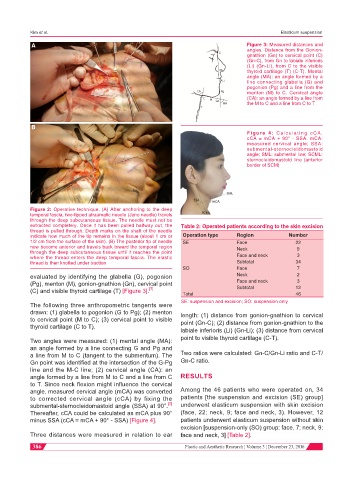
A Figure 3: Measured distances and
angles. Distance from the Gonion-
gnathion (Gn) to cervical point (C)
(Gn-C), from Gn to labiale inferioris
(Li) (Gn-Li), from C to the visible
thyroid cartilage (T) (C-T). Mental
angle (MA): an angle formed by a
line connecting glabella (G) and
pogonion (Pg) and a line from the
menton (M) to C. Cervical angle
(CA): an angle formed by a line from
the M to C and a line from C to T
B
Figure 4: Calculating cCA.
cCA = mCA + 90° - SSA. mCA:
measured cervical angle; SSA:
submental-sternocleidomastoid
angle; SML: submental line; SCML:
sternocleidomastoid line (anterior
border of SCM)
Figure 2: Operative technique. (A) After anchoring to the deep
temporal fascia, two-tipped atraumatic needle (Jano needle) travels
through the deep subcutaneous tissue. The needle must not be
extracted completely. Once it has been pulled halfway out, the Table 2: Operated patients according to the skin excision
thread is pulled through. Depth marks on the shaft of the needle
indicate how much of the tip remains in the tissue (about 1 cm or Operation type Region Number
1/2 cm from the surface of the skin). (B) The posterior tip of needle SE Face 22
now become anterior and travels back toward the temporal region Neck 9
through the deep subcutaneous tissue until it reaches the point
where the thread enters the deep temporal fascia. The elastic Face and neck 3
thread is then knotted under traction Subtotal 34
SO Face 7
evaluated by identifying the glabella (G), pogonion Neck 2
(Pg), menton (M), gonion-gnathion (Gn), cervical point Face and neck 3
[7]
(C) and visible thyroid cartilage (T) [Figure 3]. Subtotal 12
Total 46
SE: suspension and excision; SO: suspension only
The following three anthropometric tangents were
drawn: (1) glabella to pogonion (G to Pg); (2) menton length: (1) distance from gonion-gnathion to cervical
to cervical point (M to C); (3) cervical point to visible point (Gn-C); (2) distance from gonion-gnathion to the
thyroid cartilage (C to T).
labiale inferioris (Li) (Gn-Li); (3) distance from cervical
Two angles were measured: (1) mental angle (MA): point to visible thyroid cartilage (C-T).
an angle formed by a line connecting G and Pg and
a line from M to C (tangent to the submentum). The Two ratios were calculated: Gn-C/Gn-Li ratio and C-T/
Gn point was identified at the intersection of the G-Pg Gn-C ratio.
line and the M-C line; (2) cervical angle (CA): an
angle formed by a line from M to C and a line from C RESULTS
to T. Since neck flexion might influence the cervical
angle, measured cervical angle (mCA) was converted Among the 46 patients who were operated on, 34
to corrected cervical angle (cCA) by fixing the patients [the suspension and excision (SE) group]
submental-sternocleidomastoid angle (SSA) at 90°. [7] underwent elasticum suspension with skin excision
Thereafter, cCA could be calculated as mCA plus 90° (face, 22; neck, 9; face and neck, 3). However, 12
minus SSA (cCA = mCA + 90° - SSA) [Figure 4]. patients underwent elasticum suspension without skin
excision [suspension-only (SO) group: face, 7; neck, 9;
Three distances were measured in relation to ear face and neck, 3] [Table 2].
386 Plastic and Aesthetic Research ¦ Volume 3 ¦ December 23, 2016