
Page 119 - Read Online
P. 119
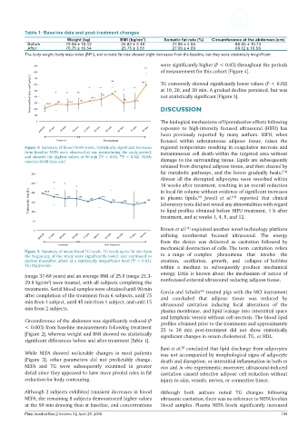
Table 1: Baseline data and post-treatment changes
                           Weight (kg)         BMI (kg/cm )     Somatic fat rate (%)  Circumference at the abdomen (cm)
                                                        2
            Before        70.95 ± 18.42         25.82 ± 3.49        27.99 ± 4.05            88.85 ± 10.73
            After         70.75 ± 18.54         25.75 ± 3.51        27.99 ± 4.09            88.12 ± 10.58
           The body weight, body mass index (BMI), and somatic fat rate showed slight decreases from the baseline, but they were statistically insignificant
                                                               were significantly higher (P < 0.05) throughout the periods
                                                               of measurement for this cohort [Figure 4].

                                                               TG conversely showed significantly lower values (P < 0.02)
                                                               at 10, 20, and 30 min. A gradual decline persisted, but was
                                                               not statistically significant [Figure 5].


                                                               DISCUSSION

                                                               The biological mechanisms of liporeductive effects following
                                                               exposure to high-intensity  focused ultrasound  (HIFU)  has
                                                               been previously reported  by many authors. HIFU, when
                                                               focused within  subcutaneous  adipose tissue,  raises  the
           Figure 4: Summary of blood NEFA levels. Statistically significant increases   regional temperature resulting in coagulative necrosis and
           from baseline NEFA were observed at any pointsduring the study period,   instantaneous cell death within the targeted area without
           and showed the highest values at 90 min (*P < 0.05, **P < 0.02). NEFA:
           nonesterifield fatty acid                           damage to the surrounding tissue. Lipids are subsequently
                                                               released from disrupted adipose tissue, and then cleared by
                                                               fat metabolic pathways, and the lesion gradually heals. [7,8]
                                                               Almost all the disrupted adipocytes were resorbed within
                                                               18 weeks after treatment, resulting in an overall reduction
                                                               in local fat volume without evidence of significant increases
                                                               in  plasma lipids.   Jewell  et al.   reported that  clinical
                                                                                           [10]
                                                                              [9]
                                                               laboratory tests did not reveal any abnormalities with regard
                                                               to lipid profiles obtained before HIFU treatment, 1 h after
                                                               treatment, and at weeks 1, 4, 8, and 12.
                                                               Brown et al.  exploited another novel technology platform
                                                                         [11]
                                                               utilizing  nonthermal focused ultrasound. The energy
                                                               from the device was delivered as cavitation followed  by
                                                               mechanical destruction of cells. The term cavitation refers
           Figure 5: Summary of mean blood TG levels. TG levels up to 30 min from
           the beginning of the study were significantly lower, and continued to   to a range of complex  phenomena  that  involve  the
           decline thereafter, albeit at a statistically insignificant level (*P < 0.02).   creation,  oscillation,  growth,  and  collapse  of bubbles
           TG: triglyceride                                    within a medium to subsequently  produce  mechanical
                                                               energy. Little is known about the mechanism of action of
           (range 37-68 years) and an average BMI of 25.8 (range 21.3-
           29.8 kg/cm ) were treated, with all subjects completing the   nonfocused external ultrasound reducing adipose tissue.
                    2
           treatments. Serial blood samples were obtained until 90 min   Garcia and Schafer  treated pigs with the MCI instrument
                                                                               [5]
           after completion of the treatment from 6 subjects, until 75   and concluded that adipose tissue was reduced  by
           min from 1 subject, until 45 min from 1 subject, and until 15   ultrasound cavitation inducing focal  alterations of the
           min from 2 subjects.                                plasma membrane, and lipid leakage into interstitial space
                                                               and lymphatic vessels without cell necrosis. The blood lipid
           Circumference of the abdomen was significantly reduced (P   profiles obtained prior to the treatments and approximately
           < 0.003) from baseline measurements following treatment   25 to  30 min  post-treatment  did not  show statistically
           [Figure 2], whereas weight and BMI showed no statistically   significant changes in serum cholesterol, TG, or HDL.
           significant differences before and after treatment [Table 1].
                                                               Bani et al.  concluded that lipid discharge from adipocytes
                                                                       [6]
           While NEFA showed  noticeable changes in most patients   was not accompanied by morphological signs of adipocyte
           [Figure  3], other parameters  did not predictably change.   death and disruption, or interstitial inflammation in both ex
           NEFA and TG were  subsequently  examined  in  greater   vivo and in vivo experiments; moreover, ultrasound-induced
           detail since they appeared to have more pivotal roles in fat   cavitation caused selective adipose cell reduction without
           reduction for body contouring.                      injury to skin, vessels, nerves, or connective tissue.

           Although 2 subjects exhibited transient decreases in blood   Although  both  authors noted TG changes  following
           NEFA, the remaining 8 subjects demonstrated higher values   ultrasonic cavitation, there was no reference to NEFA levelsin
           at the 90 min drawing than at baseline, and concentrations   blood samples.  Plasma  NEFA  levels  significantly  increased
           Plast Aesthet Res || Volume 3 || April 25, 2016                                                    109