
Page 118 - Read Online
P. 118
INTRODUCTION A paired t-test was performed for each group to determine
the presence or absence of significant differences between
Nonsurgical body contouring devices, including cryolipolysis, baseline and post-treatment for weight, body mass index
low-level laser therapy, radiofrequency instruments, and (BMI), and circumference reduction. Subjects provided written
external ultrasound devices, are widely used noninvasive informed consent prior to participation in the study.
procedures for body contouring and fat reduction. In a
clinical environment, the most effective treatments for RESULTS
noninvasive fat reduction involve ultrasound, using either
focused or nonfocused waves depending on how ultrasonic Ten subjects (5 males, 5 females) with an average age of 45
energy is delivered to the tissue. [1-4]
The MC1 instrument (General Product S.r.I., Montespertoli,
Italy) uses nonfocused ultrasound, and is designed to induce
stable cavitation while reducing adipose tissue volume in
treated tissue. [2,5,6]
Despite its apparent clinical efficacy, biological mechanisms
reducing adipose tissue are not fully understood. Adipose
cell cavitation induced focal alterations of the plasma
[5]
membrane and lipid leakage during in vivo porcine studies.
Bani et al. reported that ultrasound cavitation induced a
[6]
statistically significant reduction in the size of adipocytes,
the appearance of micropores, and triglyceride (TG) leakage.
The primary objective of this study was to document blood
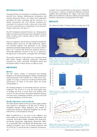
lipid profile changes following nonfocused ultrasound Figure 1: MC1 device. The MC1 device has an ultrasound and a zonal
massage handpiece. The ultrasound handpiece with angled, nonfocused
treatments for body contouring, elucidating clinical and emitters deliver 1 MHz ultrasound. In modulated emission mode, the
biochemical effects of these devices on adipose tissue. nonfocused beams create a slightly focused ultrasound field within the
tissue at the point where the beams overlap
METHODS
Device
The MC1 device consists of ultrasound and drainage
handpieces. The ultrasound handpiece delivers a dualinclined
nonfocused beam of 20 to 60 KHz on a 1 MHz carrier wave,
capable of creating a weakly focused ultrasound field within
subcutaneous fat tissue at the point where the beams
overlap [Figure 1].
The draining handpiece is used during both pre- and post-
treatments. The former is to open the main lymph nodes
to facilitate the flow of excess fluids and facilitate their Figure 2: Summary of abdominal circumference. The circumference
removal. The latter promotes immediate lymphatic drainage at the abdomen was significantly reduced (*P < 0.003) from baseline
measurements
of treated areas, stimulating metabolic processes that lead
to natural elimination of the fat waste.
Study objectives and methods
Five males and five females (37-67 years old) in supine position
were exposed to ultrasound at 20 KHz and 3.0 W/cm in
2
modulated emission. Whole abdomens were treated by
continuously moving an ultrasound handpiece for 30 min,
followed by a 6-min lymphatic drainage.
Waist circumferences at the level of the umbilicus and
body weight were measured before and immediately after
treatment. Blood lipid profiles including total cholesterol,
low-density lipoprotein, high-density lipoprotein (HDL),
TG, nonesterified fatty acid (NEFA) and lipoprotein-a were Figure 3: Blood NEFA changes in 10 subjects. Most subjects displayed
analyzed at baseline, every 10 min during treatment, and increased blood NEFA levels from the early phase and had higher values at
15, 30, 45, 60, 75, and 90 min after completion of treatment. termination of the experiments. NEFA: nonesterifield fatty acid
108 Plast Aesthet Res || Volume 3 || April 25, 2016