
Page 97 - Read Online
P. 97
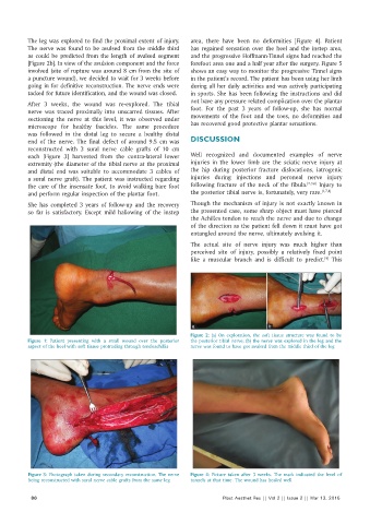
The leg was explored to find the proximal extent of injury.   area, there have been no deformities  [Figure  4]. Patient
          The  nerve  was  found  to  be  avulsed  from  the  middle  third   has  regained  sensation  over  the  heel  and the  instep  area,
          as could be predicted from the length of avulsed segment   and the progressive Hoffmann‑Tinnel signs had reached the
          [Figure 2b]. In view of the avulsion component and the force   forefoot area one and a half year after the surgery. Figure 5
          involved (site of rupture was around 8 cm from the site of   shows an easy way to monitor the progressive Tinnel signs
          a puncture wound), we decided to wait for 3 weeks before   in the patient’s record. The patient has been using her limb
          going in for definitive reconstruction. The nerve ends were   during all her daily activities and was actively participating
          tacked for future identification, and the wound was closed.  in sports. She has been following the instructions and did
                                                              not have any pressure related complication over the plantar
          After 3  weeks, the wound was re‑explored. The tibial
          nerve was traced proximally into unscarred tissues. After   foot. For the past 3  years of follow‑up, she has normal
          sectioning the nerve at this level, it was observed under   movements of the foot and the toes, no deformities and
          microscope for healthy fascicles. The same procedure   has recovered good protective plantar sensations.
          was  followed  in  the  distal  leg  to  secure  a  healthy  distal
          end of the nerve. The final defect of around 9.5  cm was   DISCUSSION
          reconstructed with 3 sural nerve cable grafts of 10  cm
          each  [Figure  3] harvested from the contra‑lateral lower   Well recognized and documented examples of nerve
          extremity (the diameter of the tibial nerve at the proximal   injuries in the lower limb are the sciatic nerve injury at
          and distal end was suitable to accommodate 3 cables of   the hip during posterior fracture dislocations, iatrogenic
          a  sural  nerve  graft).  The  patient  was  instructed  regarding   injuries  during  injections  and peroneal  nerve  injury
          the care of the insensate foot, to avoid walking bare foot   following  fracture of the neck  of the fibula. [2,3,6]  Injury to
          and perform regular inspection of the plantar foot.  the posterior tibial nerve is, fortunately, very rare. [1,7,8]
          She has completed 3  years of follow‑up and the recovery   Though the mechanism of injury is not exactly known in
          so far is satisfactory. Except mild hallowing of the instep   the presented case, some sharp object must have pierced
                                                              the Achilles tendon to reach the nerve and due to change
                                                              of the direction as the patient fell down it must have got
                                                              entangled around the nerve, ultimately avulsing it.
                                                              The actual  site of nerve injury was much higher than
                                                              perceived site of injury, possibly a relatively fixed point
                                                                                                          [9]
                                                              like a muscular branch and is difficult to predict.  This


                                                               a                       b
                                                              Figure 2:  (a) On exploration, the soft tissue structure was found to be
          Figure  1:  Patient  presenting  with a small wound over the posterior   the posterior tibial nerve; (b) the nerve was explored in the leg and the
          aspect of the heel with soft tissue protruding through tendoachillis  nerve was found to have got avulsed from the middle third of the leg


          Figure 3: Photograph taken during secondary reconstruction. The nerve   Figure 4:  Picture  taken  after  3 weeks.  The  mark indicated the  level of
          being reconstructed with sural nerve cable grafts from the same leg  tunnels at that time. The wound has healed well

            86                                                           Plast Aesthet Res || Vol 2 || Issue 2 || Mar 13, 2015