
Page 100 - Read Online
P. 100
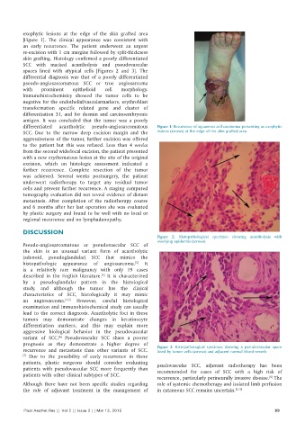
exophytic  lesions  at  the  edge  of  the  skin  grafted  area
          [Figure  1].  The  clinical  appearance  was  consistent  with
          an early recurrence. The patient underwent an urgent
          re‑excision with 1 cm margins followed by split‑thickness
          skin grafting. Histology confirmed a poorly differentiated
          SCC with marked acantholysis and pseudovascular
          spaces lined with atypical cells [Figures  2 and 3]. The
          differential  diagnosis  was  that  of  a  poorly  differentiated
          pseudo‑angiosarcomatous SCC or true angiosarcoma
          with   prominent   epithelioid  cell  morphology.
          Immunohistochemistry showed the tumor cells to be
          negative for the endothelial/vascularmarkers, erythroblast
          transformation specific related gene and cluster of
          differentiation 31, and for desmin and carcinoembryonic
          antigen. It was concluded that the tumor was a poorly
          differentiated  acantholytic  pseudo‑angiosarcomatous  Figure 1: Recurrence of squamous cell carcinoma presenting as exophytic
          SCC. Due to the narrow deep excision margin and the   lesions (arrows) at the edge of the skin grafted area
          aggressiveness of the tumor, further excision was offered
          to the patient but this was refused. Less than 4  weeks
          from the second wide/local excision, the patient presented
          with a new erythematous lesion at the site of the original
          excision, which on histologic assessment indicated a
          further recurrence. Complete resection of the tumor
          was achieved. Several weeks postsurgery, the patient
          underwent  radiotherapy  to  target  any  residual  tumor
          cells and prevent further recurrence. A staging computed
          tomography evaluation did not reveal evidence of distant
          metastasis.  After  completion  of  the  radiotherapy  course
          and 6 months after her last operation she was evaluated
          by plastic surgery and found to be well with no local or
          regional recurrence and no lymphadenopathy.

          DISCUSSION
                                                              Figure 2:  Histopathological specimen  showing  acantholysis with
                                                              overlying epidermis (arrows)
          Pseudo‑angiosarcomatous  or  pseudovascular  SCC  of
          the skin is an unusual variant form of acantholytic
          (adenoid, pseudoglandular) SCC that mimics the
          histopathologic appearance of angiosarcoma.  It
                                                      [5]
          is a relatively rare malignancy with only 19  cases
          described in the English literature.  It is characterized
                                         [4]
          by  a  pseudoglandular  pattern in  the  histological
          study,  and  although  the  tumor  has  the  clinical
          characteristics of SCC, histologically it may mimic
          an  angiosarcoma. [4,5]   However, careful  histological
          examination and immunohistochemical study can usually
          lead  to  the  correct  diagnosis.  Acantholytic  foci  in  these
          tumors may demonstrate changes in keratinocyte
          differentiation markers, and this  may explain more
          aggressive biological behavior in the pseudovascular
          variant of SCC.  Pseudovascular SCC share a poorer
                        [6]
          prognosis as they demonstrate a higher degree of
          recurrence  and  metastasis  than  other  variants  of  SCC.  Figure 3:  Histopathological specimen  showing  a pseudovascular  space
                                                              lined by tumor cells (arrows) and adjacent normal blood vessels
          [7]   Due  to  the  possibility  of  early  recurrence  in  these
          patients, plastic surgeons should consider evaluating   psudovascular  SCC, adjuvant radiotherapy has been
          patients  with  pseudovascular  SCC  more  frequently  than   recommended for cases of SCC  with  a high  risk of
          patients with other clinical subtypes of SCC.
                                                              recurrence, particularly perineurally invasive disease.  The
                                                                                                          [8]
          Although there have not been  specific studies regarding   role of systemic chemotherapy and isolated limb perfusion
          the  role  of adjuvant treatment  in  the  management  of   in cutaneous SCC remains uncertain. [9,10]


          Plast Aesthet Res || Vol 2 || Issue 2 || Mar 13, 2015                                             89