
Page 103 - Read Online
P. 103
He revealed a history of insidious onset as a small nodule,
gradually reaching the present size. The patient had no
history of trauma to this region. Extraoral examination
showed facial swelling with obliteration of left nasolabial
fold. Swelling was oval, smooth, approximately
3 cm × 3 cm in size, extending antero‑posteriorly from
left ala of nose to canine fossa and superoinferiorly from
left infraorbital foramen region to the left corner of upper
lip. The skin over the swelling appeared normal without
pain, pus discharge, or paresthesia. No lymphadenopathy
was noted.
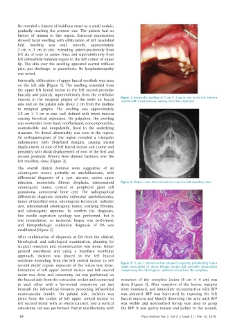
Intraorally, obliteration of upper buccal vestibule was seen
on the left side [Figure 1]. The swelling extended from
the upper left lateral incisor to the left second premolar
buccally and palataly, superoinferiorly from the vestibular
mucosa to the marginal gingiva of the teeth on buccal Figure 1: Intraorally, swelling of 3 cm × 3 cm in size in the left anterior
maxilla with intact mucosa causing bicortical expansion
side and on the palatal side about 2 cm from the midline
to marginal gingiva. The swelling was approximately
2.5 cm × 3 cm in size, well defined with intact mucosa
causing bicortical expansion. On palpation, the swelling
was nontender, bony hard, nonfluctuant, noncompressible,
nonreducible and nonpulsatile, fixed to the underlying
structure. No dental abnormality was seen in the region.
An orthopantogram of the region revealed a triangular
radiolucency with ill‑defined margins, causing mesial
displacement of root of left lateral incisor and canine and
proximity with distal displacement of root of the first and
second premolar. Water’s view showed haziness over the
left maxillary sinus [Figure 2].
The overall clinical features were suggestive of an
odontogenic tumor, probably an ameloblastoma, with
differential diagnosis of a cyst, abscess, canine space
infection, monostotic fibrous dysplasia, adenomatoid Figure 2: Water’s view showed haziness over the left maxillary sinus
odontogenic tumor, central or peripheral giant cell
granuloma, aneurysmal bone cyst. The radiographical
differential diagnosis includes unilocular ameloblastoma,
tumor of maxillary sinus, odontogenic keratocyst, radicular
cyst, adenomatoid odontogenic tumor, ossifying fibroma,
and odontogenic myxoma. To confirm the diagnosis,
fine needle aspiration cytology was performed, but it
was inconclusive, so incisional biopsy was performed,
and histopathologic evaluation diagnosis of DA was
established [Figure 3].
After confirmation of diagnosis as DA from the clinical,
histological, and radiological examination, planning for
surgical resection and reconstruction was done. Under
general anesthesia and using a maxillary vestibular
approach, incision was placed in the left buccal
vestibule extending from the left central incisor to left
second molar region, exposure of the lesion was done. Figure 3: H and E stained section showed irregularly proliferating tumor
island surrounded by dense fibrous stroma and extensive desmoplasia
Extraction of left upper central incisor and left second compressing the odontogenic epithelial island from the periphery
molar was done and osteotomy cut was performed on
the buccal side from the extraction socket and connected resection of the complete lesion (4 cm × 4 cm) was
to each other with a horizontal osteotomy cut just done [Figure 4]. After resection of the lesion, margins
beneath the infraorbital foramen protecting infraorbital were examined, and immediate reconstruction with BFP
neurovascular bundle. On palatal side, incision was was planned. BFP was harvested by exposing the left
given from the socket of left upper central incisor to buccal mucosa and bluntly dissecting the area until BFP
left second molar with an electrocautery, and a vertical was visible and nontoothed forcep was used to grasp
osteotomy cut was performed. Partial maxillectomy with the BFP. It was gently teased and pulled to the wound.
92 Plast Aesthet Res || Vol 2 || Issue 2 || Mar 13, 2015