Page 104 - Read Online
P. 104

The BFP was expanded and sutured with 4‑0 vicryl to   DISCUSSION
          the underlying  wound.  A  saframycin based  oral  pack
          was  secured  over  the  palate  with  3‑0  vicryl  suture  for   The use of BFP as a grafting  source was first described
          primary protection for 1 week till the epithelization   in 1977  by Egyedi.  Anatomically BFP is a fatty mass
                                                                               [7]
          begins,  and  strict  soft  diet  was  followed  [Figure  5].   in  the  buccal space of the cheek. It  comprises 3
          The patient was kept on periodic recall every 2 months   lobes:  anterior,  intermediate  and posterior  with  4
          during the 1 year follow‑up [Figure 6].             extensions,  that is, buccal, pterygoid, pterygopalatine
                                                              and temporal  (superficial and profound).  It is fixated by
                                                              ligaments to the maxilla, posterior zygomatic bone, inner
                                                              and outer  rim  of the  orbital  fissure,  temporalis  tendon
                                                              and buccinators membrane.  It is  intimately  associated
                                                                                      [8]
                                                              with muscles of mastication, facial nerve and parotid
                                                              duct. The  use  of  the  BFP as  a  grafting  source in  closure
                                                              of intra‑oral defects has gained popularity,  because
                                                              of the  ease  of harvesting,  simplicity,  versatility,  rich
                                                              blood supply, low complication rate and quick surgical
                                                              techniques.  Tideman  et  al.  showed that the BFP is
                                                                                       [9]
                                                                        [5]
                                                              epithelialized within 3‑4  weeks and therefore further
                                                              skin graft are not required [Figure 5]. According to Alkan
                                                              et  al.,  the  success rate  of the  use  of BFP is  relatively
                                                                   [10]
                                                              high in all comparative studies.
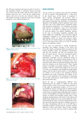
                                                              In our case, we performed a partial  maxillectomy
                                                              procedure and complete resection of the lesion was
          Figure  4:  Partial maxillectomy  was carried out and resection  of the   done, after which a postsurgical defect was present in left
          complete lesion was done
                                                              anterior maxillary region.  An immediate  reconstruction
                                                              with BFP was planned, and BFP was harvested from
                                                              the left buccal mucosa. Intra‑oral postsurgical defects
                                                              reconstruction is  always challenging  one  due to
                                                              anatomical constraints and the specialized nature of
                                                              intra oral tissues. The principal arterial supply to BFP is
                                                              from  buccal and  deep branches of maxillary  artery,  from
                                                              transverse facial branch of superficial temporal artery and
                                                                                                 [11]
                                                              from  few branches  of the  facial artery.  There  should
                                                              remain  a  reasonable  size  pedicle attached to the  BFP to
                                                              provide it  with  the  crucial blood supply in  the  1st  week
                                                              of its life. It is essential to stop bleeding from BFP during
                                                              surgery with the help of electrocautery or small ligatures
                                                              as a failure to do so leads to the formation of buccal
                                                              haematoma  and could compromise  the  viability  of the
                                                              flap. It is also important to be meticulous in dissecting
                                                              out the flap, protecting the small branches of the facial
          Figure  5:  Reconstruction with buccal pad  of fat. Immediate   nerve and parotid duct.
          intra‑operative picture
                                                              Buccal fat  pad is  morphologically different  from
                                                              subcutaneous fat but similar to orbital fat. The mean
                                                              volume of BFP is  about 10  mL  and its  mean  thickness is
                                                              6  mm  while  the  approximate  weight  is  of 9.3  g.  It  can
                                                              successfully be used for covering small to medium defects
                                                                                     [12]
                                                              of about 4 cm in diameter.  BFP has also been employed
                                                              in the closure of surgical defects following tumor excision,
                                                              excision of leukoplakia and submucous fibrosis, as well as
                                                                                                         [13]
                                                              closure  of primary  and secondary  palatal clefts.   Flap
                                                              should be sutured gently to the borders of the defects and
                                                              ideally there should not be any stretch within the tissue.
                                                              Overstretching the tissue can lead to fragmentation of the
                                                              flap and in the long term can lead to ischemic necrosis at
                                                              the  edges.  Disadvantage with  the  use  of the  BFP flap is
                                                              hematoma  formation,  partial necrosis,  excessive  scarring,
                                                              infection or facial nerve injury.  None of these  changes
          Figure 6: Postoperative picture after 1 year        was noted in our case. The use of BFP in patients with
          Plast Aesthet Res || Vol 2 || Issue 2 || Mar 13, 2015                                             93
   99   100   101   102   103   104   105   106   107   108   109